The skeleton is made up of bones with the main roles of support and protection, as well as allowing the movement of vertebrate animals. The skeletal muscles are usually associated with bones but also with cartilage and are sometimes free muscles. Skeletal muscles are attached to bones through tendons. The movement is produced by muscle contraction and relaxation. We are going to include bones, tendons, and skeletal muscles in the motor system.
Skeleton
The skeleton consists of bones (Figure 1). It performs three main functions: supporting body structures, protecting vital organs, and forming stiff structures (long or short bones) that the muscles can move in order to move parts of the body or commute the whole body. The skeleton is also a store for calcium minerals, blood cells, and an energy reservoir in the form of fat in adipocytes.

Mechanical support. The skeleton is a scaffold for supporting and organizing the body's structures and organs. Without the skeleton, the rest of the body would collapse like soft dough. For instance, the brain is a very soft tissue that would spill out outside the skull. The viscera are encased by the thoracic compartment and supported by the spine. Muscles, although more consistent, also keep their position and organization through attachment to the bones. The shape and size of our body largely depend on our bones. For instance, consider the length of the limbs and their components.
Protection. The animal body contains many delicate organs, such as the nervous system (encephalon and spinal cord), the lungs, and the heart. They need to be protected against strokes and mechanical insults. The nervous system is protected by the skull and vertebra, whereas the ribs and breastbone protect the lungs and the heart.
Movement. The coordinated action of muscles and bones in our limbs allows us to go from one place to another. They are also necessary for the movement of body parts, like the head, fingers, or postural changes. The bones act as levers connected through tendons to the muscles, which provide the power for movement.
Calcium storage. Calcium is found in the extracellular matrix of bones as hydroxyapatite minerals. When the mineral is solved, the calcium is freed and can enter the bloodstream to reach whatever part of the body.
Blood cell production. In the spongy bone, many hematopoietic cells are found in the bone marrow. They are stem cells that proliferate and differentiate into blood cells. The bone marrow includes the hematopoietic stem cells as well as all cell types found in the bone cavities.
There are 206 bones in the human body (300 in babies, where some bones are fused during development). Bones are living structures with blood vessels and nerves, and continuous renewing is carried out by bone cells: osteoblasts, osteoclasts, and osteocytes. As mentioned before, many cell types are found in the bone marrow. A typical bone consists of an outer layer of compact bone, where the spongy bone is found within the bone structure.
Bones can be classified in different ways. Regarding their position, there are axial and appendicular bones. The axial skeleton is found along the middle line of the body, and it is composed of 80 bones that include the skull, the hyoid bone, auditory bones, ribs, the breastbone, and vertebrae. The appendicular skeleton is formed of the fore and hind limbs, pelvis, and shoulders. According to their shape, the bones are classified into large, short, flat, irregular, sesamoids, and sutural bones.
Muscles
Muscles provide the power for body movements or to maintain position. There are about 650 muscles in the human body (Figure 2). Together with bones, muscles give shape to the body and are about half of the total weight of the body. Most muscles that allow movement are composed of striated skeletal muscle cells, connective tissue, blood vessels, and nerves, connected to the bone through tendons, although some muscles are directly attached to the bone. Some muscles, such as the tongue and some muscles of the face for facial expressions, produce movement without being connected to any bone. The digestive tract is moved, and the diameter of the arteries changes with the contraction of smooth muscle cells. In the same way, the heartbeat is an independent movement produced by the cardiac muscle cells. We will deal with the muscles associated with bones.

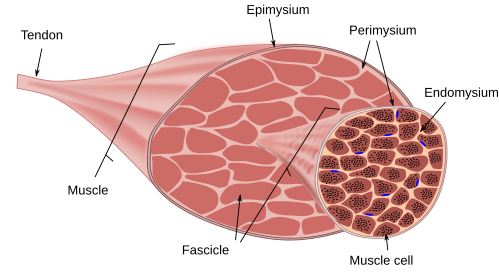
The skeletal striated muscle cells form the skeletal muscle of our body. They are also called muscle fibers or myocytes. Muscles are also made up of blood vessels, nerves, and connective tissue. Muscle cells are packaged into fascicles, and several fascicles form a muscle (Figure 3). Muscle cells are wrapped by a layer of connective tissue called the endomysium; each fascicle is covered by the perimysium, which is a layer of dense connective tissue; and the epimysium is the connective sheath that covers the whole muscle. The blood vessels and nerve fibers enter the muscle and branch many times through these connective layers. The nerves control the muscle contraction.

Skeletal muscles produce voluntary movement by shortening the muscle cells. The many muscles that form the animal body can be named according to their position. The axial muscles are those related to the spine and tail, and the appendicular muscles are involved in limb movements. A few muscles are the branchyomeric muscles, which are related to the jaws and the hyoid apparatus.
Tendons
Tendons connect muscles with bones, so they are intermediaries between these two components of the motor system. Tendons are also involved in stabilizing joints, such as in the knees and the back. Regular, dense connective tissue is the main component of tendons. The fibers of the extracellular matrix, mostly thick collagen fibers, are oriented parallel to the strength axis, which confers great resistance to stretching.
Tendons are largely dense, regular connective tissue with thick bundles of collagen fibers and cells known as tenocytes. They are covered by a sheath of connective tissue called the endotenon. This layer is in contact with the dense, collagen bundles and sends inward walls of tissue that carry blood vessels and nerves. There is an outer layer, known as the epitenon, which covers the whole tendon surface. Some tendons show additional layers, such as paratenon. The outer layer facilitates the movement of the tendon and friction with other tissues.
The attachment point of the tendon to the bone is known as the myotendinous junction, which is a complex structure. At the end of the skeletal muscle cells, the plasma membrane deeply folds, and tendon collagen bundles are inserted in the invaginations. On the bone side, the connective tissue of the tendon is among the bone matrix, with a transition of fibrous cartilage that is progressively mineralized toward the bone. The tendon collagen fibers enter the bone by crossing the periosteum, forming the so-called Sharpey's fibers. Macroscopically, the bone surface is rough, with small pores at the insertion point.
Ligaments are dense connective tissue that join bones to each other at the joints. They are fibrous layers that attach their ends to close bones, therefore maintaining the structural integrity of joints. However, the so-called yellow ligaments are composed of elastic fibers and connect the upper parts of the vertebral arch.
Joints
Bones are associated with each other at joints to produce body movements. Cartilage, ligaments, and lubricant liquid are also joint components. According to movement capability, there are several types of joints.
Joints are named after the bones, or part of the bones, that form the joint. For instance, the radioulnar joint, the temporomandibular joint, the femorotibial joint, and so on. Structurally, they are classified considering the tissue more abundant in the joint: fibrous, cartilaginous, and synovial joint. Functionally, there are joints where bones are not able to move (synarthrosis), those that can move slightly (amphiarthrosis), and those with high mobility (diarthrosis). In the anatomy books, the structural classification is found, but the functional names are used, including those joints with low mobility included in the synarthrosis type. We will follow the functional names.
Synarthrosis
Since movement is not the main function of synarthrosis, the tissues that form this type of joint are mostly connective and cartilaginous tissues. Its function is to keep together parts of the skeleton or give support to the growth of the skeletal components involved in the joint.
Syndemosis is a type of synarthrosis that has connective tissue connecting the bones. Ligaments (dense connective tissue) maintain the cohesion of these joints. The sutures between the skull bones are examples of syndemosis. In newborns, there is a membrane called the fontanelle that allows for higher flexibility of the bones during the growth of the head. Later, the fontanelle is closed, and the connective tissue of the sutures becomes ossified. In this case, it is renamed synostosis. Other examples of syndemosis are some large bones, such as the humeroradial joint, which allows a short movement between the radius and the humerus, and the attachment of teeth to the alveoli of the jaws by the periodontal ligament, which allows a slight movement of teeth during jawing.
Synchondrosis contains hyaline cartilage between the bones. The mobility of bones in these joints is slight, or not allowed at all. This type of joint may be transient, as in the epiphyseal plates between the epiphysis and diaphysis, which allow the growth of long bones. The epiphyseal plates are ossified during puberty, and they are then called synostosis. Another example are the joints between the ribs and the breastbone. These joints allow a slight movement between the ribs and the breastbone. However, during development, only the attachment between the first rib and breastbone remains. The rest, although maintaining the cartilage, are transformed into synovial joints to allow movement during breathing.
In the symphyseal joints, the cartilage binding the bones is fibrocartilage. Examples are the intervertebral discs and the attachment of the pubis bones. There is some mobility due to the flexibility of the fibrocartilage.
Diarthorosis
Diarthrosis are the "real" joints, also known as synovial joints. The bones are separated from each other, allowing movement. The surface of the bones in the joint is covered by the articular cartilage, a type of hyaline cartilage. There is no physical contact between the surfaces of the bones since there is synovial liquid between them that fills the joint cavity. Fibrocartilage is found in some of these joints, among the articular cartilage, forming joint discs, meniscus, and joint labrums. Examples of articular disks are the temporomandibular and the sternoclavicular joints. The disks separate the joint cavity into two independent cavities. Fibrocartilage does not form closed structures in the meniscus of the knee and elbow joints. The function of the disks and meniscus is to cushion mechanical loads. Labrums are found in the shoulder and hip joints, with the role of widening the articular surface for protection against fractures.
 Endocrine