1. Introduction
Nowadays, we agree that all living organisms are made up of cells. The cell size is smaller than the resolving power of the human eye. Therefore, to observe cells, microscopes had to be invented. But even with the aid of microscopes, it was difficult to realize that cells are the units that form all living beings. This is in part a consequence of the wide diversity of morphologies and sizes that cells show.
The history of the cell discovery begins with the first lens and microscopes were made, in the early seventeenth century. The modern idea of cells, as well as its evolution over the years, is tightly linked to the crafting and refinement of microscopes, and therefore, o technology.
2. XVII century
1590-1600. A. H. Lippershey, Z. Janssen y H. Janssen (father and son). They are reckoned as the inventors of the compound microscope, that is, two lenses, each placed at each end of a tube.
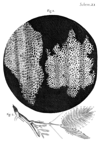
1664. R. Hooke publishes Micrographia, where he writes the first description of a cell. He was studying sheets of cork, described them as a bee honeycomb, and named "cell" to each of the little chambers (Figure 1). The name "cell" was latter applied to the content of these chambers, and was latter used to name the units of animal tissues.

1670-1680. N. Grew y M. Malpighi found the same structures, i.e. the cells, in many other plants. N. Grew first used the term "parenchyma" in plants and M. Malpighi gave names to many plant microscopic structures.
1670. A. van Leeuwenhoek crafted simple microscopes, consisting of only one lens, but so well-polished that allowed to reach 270 magnifications. He can be regarded as the father of Microbiology, since he wrote the first descriptions of prokaryote and unicellular eukaryotes. He described the features of many biological samples with unprecedented detail.
3. XVIII century
Over the eighteenth century, much progress was achieved in the polishing of lenses. In this way, microscopes provided sharper images of biological samples.
1759. The first attempt to place animal and plants at the same level was done by C. F. Wolf. He claimed that there is a globular fundamental unit that forms all living organisms. He also proposed that living organisms get the adult forms by progressive development, and the different body structures develop from others less complex structures by growing and differentiation. This idea was opposite to the preformacionist theory, very popular at that time. The preformationist theory posed that the organisms were already completely formed as tiny bodies inside the gametes, and the adult forms were achieved by just enlargement of each of the body structures.
4. XIX century
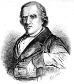
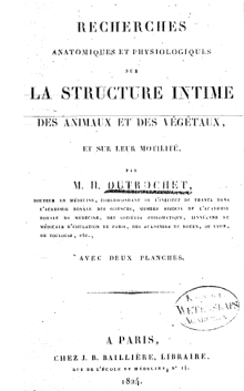
1820-1830. The outset of the cell theory starts in France. H. Milne-Edwards and F. V. Raspail (Figures 2 and 3) published that tissues were made up of globular units with uneven distribution. R. J. Dutrochet, French too, wrote (Figure 4) that if someone compares the extreme simplicity of this chocking structures, the cell, with its extremely complex content, it is clear that the cell is the basic unit of every organized organism, actually, everything is derived from the cell. R. J. Dutrochet was the first to assign to the cell a physiological role. F. V. Raspail proposed that each cell was like a laboratory, that allows the organization and function of tissues and organisms. He said, and not R. Virchow, "Omnis cellula e cellula", that is, every cell comes from other cell.


1831. R. Brown described the nucleus. A. van Leeuwenhoek, in 1682, and F. Bauer, in 1802, had been described a cellular structure that could not be anything else but the nucleus.
1832. B. Dumortier described the binary division in plant cells.
cell division
1837. J. E. Purkinje posed the basic concepts of the cell theory. He claimed that the animal tissues can be compared to plant tissues.
1838. M. J. Schleiden proposed the first tenet of the cell theory for plants: all plants are formed by basic units known as cells. T. Schwann extended this tenet to all living beings. There are other scientists that reached the same conclusion: L. Oken (1805), R. J. H. Dutrochet (1824), J. E. Purkinje (1834) y G. G. Valentin (1834).
1839-1843. F. J. F. Meyen, F. Dujardin y M. Barry connected and unified different fields of biology after showing that protozoa were individual nucleated cells, similar to those found in plant and animals.
1839-1846. J. E. Purkinje y H. van Mohl, independently named protoplasm to the interior of the cell, nucleus excluded.
1856. R. Virchow proposed the cell as the simplest living form, and, even so, the the cell completely represents the idea of life. The cell is the organic unit, the indivisible living unit.
1858. The use of dyes to study biological samples brought an unprecedented advance for distinguishing cell and tissular structures.
1899. C. E. Overton proposed a lipidic nature for the interface between the cell (protoplasm) and the environment.
5. XX century
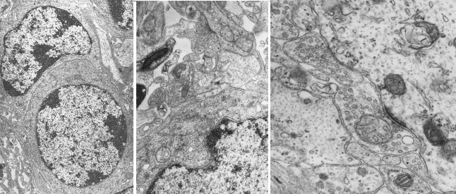
1932. The electron microscope is invented. With this apparatus, very tiny structures could be visualized (Figure 5), about nanometers (10-3 micrometers) in size. One major achievement was the visualization of membranes as a universal component of the cell. A cell delimiting membrane, the plasma membrane, was finally observed. The eukaryote cell appeared highly complex and rich in compartments.

-
Bibliography ↷
-
Bibliography
Cavalier-Smith, T. 2010. Deep phylogeny, ancestral groups and the four ages of life. Philosophical transactions of the Royal Society B. 365: 111-132
Harris, H. 2000. The birth of the cell. Yale University Prerss. ISBN-10: 0300082959
Hook, R. 1664. Micrographia. US National Library of Medicine
Ling, G. 2007. History of the membrane (pump) theory of the living cell from its beginning in mid-19th century to its disproof 45 years ago - though still taught worldwide today as established truth. Physiological chemistry and physics and medical NMR 39: 1–67.
Liu, D. 2017. The cell and protoplasm as container, object, and substance, 1835–1861. Journal of the history of biology. ) 50:889–925.
http://micro.magnet.fsu.edu/index.html
-
 Diversity