Animal organs. Central nervous system.
SPINAL CORD

Species: rat (Rattus norvegicus). Mammal.
Technique: paraffin, haematoxylin-eosin.
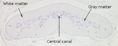
The main regions of the spinal cord can be observed in this figure, a transverse section of rat spinal cord after a general staining. The central canal is located in the medial and central part of the spinal cord. The cerebrospinal fluid flows through this canal, which is limited by the ependyma. The gray matter surrounds the central canal. Most of the somata of the spinal cord neurons localize in the gray matter, which can be subdivided in regions. Gray matter has a shape that resembles open butterfly wings, although in other species may have a more rounded shape, or ever flattened as in lampreys and hagfish (see image on the right). White matter wraps gray matter. White matter is mainly composed of axons coming from the encephalon, from the sensory neurons of the peripheric nervous system, and from the neurons located inside of the spinal cord (propriospinal neurons). Axons coming from the same source are usually packaged in bundles or funiculi located in specific positions in the white matter.

