

1. Organization
2. Development
3. Ventricles
4. Meninges
5. Hematoencephalic barrier
1. General organization
The central nervous system of vertebrates is composed of the encephalon (generally referred to as the brain) and the spinal cord (Figures 1 and 2). The encephalon is found in the head, protected by the skull, and the spinal cord extends from the encephalon until the lumbar region, encased in the spine. The encephalon is divided into three main compartments: from rostral to caudal are the primary prosencephalon, mesencephalon, and rombencephalon. The primary prosencephalon is subdivided into the secondary prosencephalon and the diencephalon. The secondary prosencephalon encompasses the telencephalon and the hypothalamus. These main compartments have been conserved during evolution in all vertebrate species studied so far. The spinal cord shows a relatively simple organization, divided into segments, with the spinal nerves at the border between adjoining segments.


2. Embryonary development
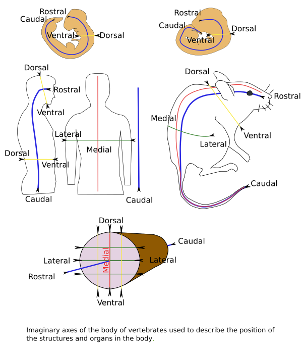
During embryonic development, the encephalon and the spinal cord derive from a group of cells known as the neural plate, located at the medial and dorsal ectoderm of the embryo (see this figure for the names of the body axes). The neural plate extends rostro-caudally. Induced by the axial mesoderm, the lateral margins of the neural plate move upward, forming long folds, whereas the middle part moves inward, forming a long groove. As the development proceeds, lateral folds, known as neural crests, get higher and closer until they contact and fuse with each other, so that a long tube is formed inside the embryo. This tube is known as the neural tube, and the process is called primary neurulation (Figure 3). During the fusion of the folds, the cells at the lateral margin of the neural plate detach from the ectoderm and set free into the embryo. These cells are known as the neuronal crests, and they migrate to many parts of the embryo to differentiate into many cell types, including the peripheral nervous system. At the most caudal part of the embryo, a tube is formed from mesenchymal tissue that first forms a row and then a hollow tube that gets fused with the caudal part of the neural tube. This process is called secondary neurulation.

The encephalon appears in the most rostral part of the neural tube. During the early stages of development, it is divided into three compartments (from rostral to caudal): primary prosencephalon, mesencephalon, and rhombencephalon (Figure 4). The caudal part of the rhombecephalon continues with the spinal cord, which extends until the caudal part of the embryo. Later in development, the primary prosencephalon is divided into two new compartments: the secondary prosencephalon and the diencephalon. The telencephalon and the hypothalamus are components of the secondary prosencephalon. The rhombencephalon has been traditionally divided into metencephalon (pons with the cerebellum) and milenecephalon (medulla oblongata), but now it is divided into rhombomeres, which are transversal segments of the rhombencephalon. These compartments have been found in the central nervous system of all vertebrates..

During embryonic development, the central nervous system is divided into a basal plate (ventral) and an alar (dorsal) plate, separated by a long and shallow groove known as the sulcus limitan of His, or sulcus limitans. Thus, each major compartment of the central nervous system shows its lateral walls divided into a basal plate and an alar plate (Figure 5). In the most rostral compartment, the pallium, subpallium, and part of the dorsal hypothalamus form the alar plate, whereas the ventral part of the hypothalamus is the basal plate. All the following segments show both the basal and the alar plates, including the spinal cord. At the midline of the ventral part of each segment, the floor plate is developed, while at the dorsal midline, the roof plate develops. In the following pages, we will deal with each of these segments, from caudal to rostral. It is worth noting that all these segments are connected and coordinated.

The production of new neurons is referred to as neurogenesis. Most neurons are produced during the embryonic and perinatal periods in vertebrates. However, neurogenesis is also observed in adult brains. In fish and reptiles, many neurons are generated during the adult period in the central nervous system. In mammals, neurogenesis is restricted to a few areas in the adult encephalon, and the number of new neurons is very low. In humans, it has also been suggested that there is limited neurogenesis in adults, with neurons migrating locally, near the production centers.
3. Ventricles / choroid plexus
The encephalon and spinal cord derive from the hollow neural tube. So, they have internal cavities that are connected to each other. These cavities are filled with cerebrospinal fluid. The largest cavities are known as ventricles, which are found in the encephalon (Figure 6), whereas a long duct, known as the central canal, or ependimary canal, runs along the spinal cord. Some regions of the roof plate of the telencephalon are not nervous tissue but epithelial tissue known as the choroid plexus (Figure 6), which is responsible for the secretion of the cerebrospinal fluid.

4. Meninges
The encephalon is caged by the skull, whereas the spinal cord is protected by the vertebral column. Meninges are thin layers of tissue found between the bones and the nervous tissue. Three meninges are described: dura mater (near the bone), arachnoid (middle), and pia mater (near the nervous tissue) (Figure 7).

The dura mater is the most superficial, thick, and resistant meninge. It is almost fused with the bone. It consists of two sheaths: a periosteal layer attached to the bone and an inner meningeal layer. The arachnoid is clear and shows many trabecules and membranous septa between the outer layer adhered to the dura mater and the deeper layer in contact with the pia mater. The inner space of the arachnoid is variable in thickness and filled with cerebrospinal fluid. It is called the subarachonoid space. The pia mater is the thinnest and deepest meninge. It covers the central nervous system and is involved in the formation of the hematoencephalic barrier.
5. Hematoencephalic barrier
About 600 km of blood vessels are estimated to be present in the encephalon. Most of them are capillaries. The blood system and the supply of oxygen are essential for the functioning of the encephalon. However, there is a very selective filtering of molecules crossing from the blood to the interneuronal space. The hematoencephalic barrier is the interface between the nervous tissue and the blood stream and filters the molecules that are exchanged between them. It consists of a layer of endothelial cells, wrapped by pericytes and astrocyte processes (Figure 8). The three elements form the neurovascular unit. The microglia, another type of glia, may collaborate to seal the barrier when an endothelial cell dies or is damaged.

The restriction to the free diffusion of the hematoencephalic barrier strongly isolates the nervous tissue from toxic substances and pathogens, but also from the immune system. Actually, it is one of the main obstacles to delivering drugs to the encephalon. Breakages of the hematoencephalic barrier may be related to epilepsy and multiple sclerosis.
-
Bibliography ↷
-
Nieuwenhuys R, Voogd J, van Huijzen Ch. El sistema nervioso central humano. 2009. 4ª Edición. Editorial Médica Panamericana S.A. ISBN: 978-84-7903-453-5.
Puelles L, Martínez S, Martínez de la Torre M. Neuroanatomía. 2008. Editorial Médica Panamericana S.A. ISBN: 978-84-7903-453-5.
-
 Nervous system