The spinal cord is the most caudal part of the central nervous system of vertebrates. It starts where the rhombencephalon ends. In humans, the spinal cord is about 2% of the total central nervous system. However, this percentage is higher in less cephalized vertebrate species. In general, the spinal cord is an intermediary structure between the encephalon and the body musculature, although many motor circuits are circumscribed to the spinal cord. The spinal cord surface is covered by layers called meninges and protected by the vertebral bones that have cavities through which the spinal cord is extended. These vertebral cavities are lined up and form a long canal known as the medullary canal.
1. Morphology
The spinal cord is elongated, and in humans, it is 1 to 1.5 cm in diameter. The shape is rather homogenous along the rostro-caudal axis, except for the most caudal part, where the diameter progressively decreases. In tetrapods, there are two thickenings found at the levels of the spinal cord segments that innervate the forelimbs (rostral) and hindlimbs (caudal), respectively. In transversal view, the spinal cord is rounded in most vertebrates. However, in some fish, such as lampreys, it is dorsoventrally flattened. Within the spinal cord, there is a duct in the central region known as the central canal, or ependimary canal, which is filled with cerebrospinal fluid and extends from the encephalon to the most caudal part of the spinal cord.
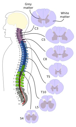
In some animals, like humans, the spinal cord is shorter than the medullary canal. In humans, the caudal part of the spinal cord ends at the level of the more rostral lumbar vertebrae (Figure 1). Some differences exist depending on the height of the individuals, so that the spinal cord ends at higher rostral levels in taller individuals. It is because vertebrae grow proportionally larger than the spinal cord. In humans, the spinal cord is 40 to 50 cm in length. The filum terminale is a non-nervous structure that anchors the spinal cord to the sacral bone. In birds and reptiles, however, the spinal cord occupies all the rostro-caudal extension of the medullary canal and does not show filum terminale. In humans, during the third month of development, the spinal cord extends all along the medullary canal, and both the spinal cord and the medullary canal grow at the same rate, but the medullary canal grows faster in later developmental stages. However, the nerves emerge from the spinal cord during the early developmental stages. This is determined by the interactions between the central nervous system and the somites. It means that the caudal levels of the spinal cord send and receive nerves that enter and exit through the caudal spine.

2. Nerves
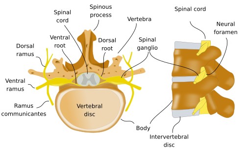
Tundles of axons are found on both sides (bilaterally) and at regular distances along the spinal cord. There are dorsal bundles (posterior), known as dorsal roots, and ventral bundles (anterior), or ventral roots. A segment is the spinal cord that extends between two consecutive bundles (Figure 2). Thus, dorsal and ventral roots are found bilaterally and at the same rostrocaudal level in each segemnt. The dorsal and ventral roots of the same side, at the same level, are fused to form the spinal nerves.

The spinal nerves leave the spine through intervertebral spaces known as intervertebral, or neural, foramens (Figure 3). Ventral roots are made up of axons that transmit motor information to the muscles. Dorsal roots bring sensory information from the body. Thus, the spinal nerves are both motor and sensory nerves, that is, they are mixed nerves. Some nerves of the sympathetic (at the thoracic to lumbar levels: T1 to L3) and parasympathetic (at the sacral level:S2-S4) preganglionic neurons, belonging to the autonomous (vegetative) system, enter the spinal cord through the ventral roots.

In humans, there are 31 pairs of the spinal nerves, named and numbered according to the vertebrae through which they get out. Thus, there are cervical (C), thoracic (T), lumbar (L), and sacral-coccygeal (S) (Figure 4). In humans, there are 8 cervical (C1-C8), 12 thoracic (T1-T12), 5 lumbar (L1-L5), 5 sacral (S1-S5) and 1 coccygeal nerves. In those species where the spinal cord does not reach the end of the medullary canal, the medullary canal without spinal cord is occupied by the nerves that exit in the caudal levesl of the spine, forming a budle of fibers known as the cauda equina (horse tail), which is absent in avian and reptiles.

3. Inner organization
In transverse view, perpendicular to the rostrocaudal axis, the spinal cord consists of two regions: grey matter and white matter (Figure 5). The names are because of the colors that these regions show in fresh tissue. The grey matter occupies a central position, and it contains most of the spinal neuronal somata, whereas the white matter wraps the grey matter and mainly contains neuronal processes, mostly propriospinal axons (axons belonging to spinal neurons that do not leave the spinal cord), spino-encephalic axons (ascending to the encephalon), and encephalo-spinal axons (descending from the encephalon). The central canal, or ependimary canal, is located in the central area of the gray matter.

Grey matter
In most vertebrates, the grey matter in transverse view looks like a butterfly with spread wings. If the central canal is taken as the central part, the grey matter can be divided into a dorsal part and a ventral part. The dorsal expansions are known as dorsal horns, and the ventral expansions are known as ventral horns (Figure 5). Both sides of the spinal cord are connected by axonal tracts, or commissures, that cross the midline, either above or below the central canal. Neurons involved in the reception and processing of sensory information are found in the dorsal horns, and from here, axons are sent to the encephalon. Motoneurons are located in the ventral horns. They form the effector motor component that innervates the voluntary muscles. The region between the dorsal and the ventral region in called intermediate, where interneurons are abundant. The region around the central canal is known as the central zone or periependimary region. In some species, like humans, there are lateral expansions of the grey matter called lateral zones. In the lateral and intermediate zones of the spinal cord, there are neurons of the autonomous system that innervate the viscera and the organs of the pelvis. The different regions of the spinal cord are also known as columns (dorsal, intermediate, lateral, and ventral) because they extend along the rostrocaudal axis of the spinal cord.

The neurons of the grey matter may also be classified into nuclei or layers (Figure 6). From the dorsal to the ventral parts, the following nuclei can be found: nucleus of the marginal zone, substantia gelatinosa, nucleus proprius, nucleus dorsalis of Clark, intermediate nuclei, and ventral motor nuclei. The grey matter can also be subdivided into layers that are named from I (dorsal) to X (ventral).

The neurons in the spinal cord can be classified according to their function. Neurons in the dorsal horn are involved in processing sensory information, and their axons form the tracts of the white matter that do not leave the central nervous system. In the ventral and lateral horns, there are motor neurons with axons that exit the spinal cord through the ventral roots. All along the the grey matter, there are propriospinal neurons, or interneurons, that form local circuits with axons that do not leave the spinal cord. The propriospinal neurons may be up to 90 % of the total neurons of the spinal cord.
The primary sensory inputs arrive at the dorsal horn, coming from the skin and inner tissues of the central body and limbs. These nerves are sent by different types of neurons. Nociceptive information ends mainly in the I, II, and V layers of the dorsal horn. Much information arrives at the dorsal horn from different parts of the body. This information has to be processed according to other contextual information and is carried out largely by the interneurons, which are up to 99 % of the total dorsal horn neurons. There are excitatory and inhibitory interneurons, which can be subdivided into more types depending on the neurotransmitter they express.
The central canal runs along the spinal cord, and it is continuous rostrally with encephalic cavities. It is lined by a layer of ependimary cells. The cerebrospinal fluid runs through the central canal.
White matter
The white matter is divided into bundles (called funiculi or columns) of nervous fibers that run parallel to the rostrocaudal axis (Figure 6). Each funiculus is composed of axons. Most of these axons are myelinated. The different bundles are characterized by the type of information they carry along the spinal cord. Depending on the location of the neuronal somata that send these axons, they are descending bundles when the soma is located in the encephalon, ascending bundles when the soma is located outside the encephalon and the axons reach the encephalon, or propriospinal when both soma and axon are confined to the spinal cord and communicate different levels of the spinal cord. There are three main tracts: the dorsal column or medial lemniscus (ascendant), the spinothalamic tract (ascendant), and the corticospinal tract (descendant). Each fiber tract is positioned in a specific place of the white matter. For instance, axon bundles coming from the cerebellum are located laterally, axons from cortical areas are located more dorso-medially, axon tracts from the thalamus run through the medio-ventral area, and ascending sensory information travels along axon bundles located dorsally.
The ventral commissure of the spinal cord courses below the central canal and carries information either from or to the ecephalon. This commissure is found along the spinal cord, and it is a fundamental structure to communicate one side of the encephalon with the contralateral side (the other side) of the body, carrying both sensory and motor information.
-
Bibliography ↷
-
Bibliography
Puelles L, Martínez S, Martínez de la Torre M. 2008. Neuroanatomía. Editorial Médica Panamericana S.A. ISBN: 978-84-7903-453-5.
-
 Central nervous systems