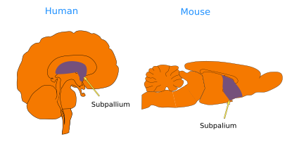
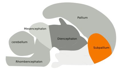
The subpallium is found ventrally to the pallium, and both form the telencephalon (Figures 1 and 2). Unlike the pallium, which is formed mainly by neurons arranged in layers, the subpallium contains neurons organized in nuclei, or groups. The subpallium encompasses the striatum, globus pallidus, area innominata, preoptic region, and ventral septum, as well as some parts of the amygdala. All these structures have been traditionally related to motor control, but it is now acknowledged their role in other functions, such as emotions, motivation, and thinking, that may lead to movement. Therefore, they are not just involved in sensorimotor circuits.


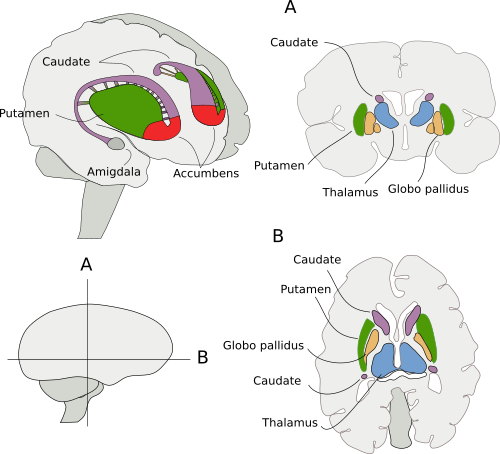
The striatum is divided into the accumbens, caudate, and putamen nuclei (Figure 3). It is the largest structure of the subpallium, and it is involved in the control of motor abilities and voluntary movements, as well as other functions. This is clear in Parkinson disease, when the striatum loses the dopaminergic afferent, leading to malfunctions and failures of motor behavior and normal cognitive activity. Cortical regions, such as the sensorimotor cortex, association cortical areas, and limbic areas, send direct inputs to the striatum. There are also afferents from the thalamus and mesencephalic dopaminergic populations. The information processed by the striatum is largely sent to the globus pallidus and ventral pallidus.

The striatum is organized into the so-called striosomes and the matrix. Both compartments contain projection GABAergic neurons. The striosomes are regions with low acetylcholinesterase activity and high levels of µ opioid receptor (MOR) expression. They appear early during embryonic development and receive the first cortical and mesencephalic dopaminergic afferents.
The globus pallidus, together with the striatum, form the basal ganglia. It is involved in motor activity. In addition, the globus pallidus, the striatum, and the dopaminergic afferent from the substantia nigra are involved in the addiction and reward mechanisms.
Several structures are found in the substantia innominata, such as the basal nucleus of Meynert, the diagonal band of Broca, and the interstitial horizontal nucleus. These nuclei, together with the septum, provide most of the cholinergic innervation to the cortical areas, releasing the acetylcholine neurotransmitter.
The preoptic region is found in the most rostral part of the third ventricle. It encompasses the median preoptic nucleus, the periventricular preoptic nucleus, and the medial preoptic nucleus. There are neurons arranged in a rather reticular organization in the lateral preoptic region.
Functions
Performing precise body movements in a specific sequence is ruled by cortical and subcortical projections to the striatum. These afferents synapse on GABAergic striatal neurons. GABAergic neurons may make up 90–95% of the total striatal neurons. From the striatum, there is a direct projection to the basal ganglia and external globus pallidus. These GABAergic neurons express the dopamine receptor D1 and the substance P neurotransmitter. Other GABAergic neurons form the indirect pathway by sending axons to the external globus pallidus, which in turn sends axons to the basal projection nuclei (that is why the name is indirect). These GABAergic neurons express the dopamine receptor D2 and the enkephalin neurotransmitter. The direct pathway promotes the disinhibition of the thalamus and the beginning of motor activity, whereas the indirect pathway does the opposite.
-
Bibliography ↷
-
Bibliography
Puelles L, Martínez S, Martínez de la Torre M. Neuroanatomía. 2008. Editorial Médica Panamericana S.A. ISBN: 978-84-7903-453-5.
-
 Hypothalamus