1. Features
2. Hyalin cartilage
- Articular cartilage
3. Elastic cartilage
4. Fibrous cartilage
Together with bones, cartilage is one of the main supporting tissues in animals. This function mainly relies on the extracellular matrix. Cartilage is a semi-rigid structure that maintains the shape of several organs, covers the surface of bones in the joints, and is the main supporting tissue during embryonic development, when bones are not yet present. During development, bone substitutes cartilage by endochondral ossification. During evolution, cartilage was the tissue that allowed the formation of vertebrate endoskeleton. Most cartilage of vertebrates differentiates from mesoderm. However, some cartilages, like craneofacial cartilage, is derived from neural crests. There are tissues called chondroid that show intermediate features between cartilage and bone.
1. Features
Ccartilage is mostly an avascular tissue, lacking blood and lymphatic vessels, and without nerve terminals. The mechanical and biochemical features of cartilage depend on the extracellular matrix, which is mainly composed of collagen (15-20 %; type II collagen being the mos abundant), proteoglycans (mainly aggrecan) and glycoproteins (10 %), and water (65-80%). Long molecules of hyaluronan are also present in the cartilage extracellular matrix. Collagen is important for withstanding stretching, while aggrecan resists mechanical pressures and provides abundant hydration. Extracellular matrix may be mineralized or non mineralized. Although cartilage is avascular, there are channels called cartilaginous channels in the embryonary cartilage that bring blood vessels for short distances into the cartilage. These channels also allow the entrance of chondroblasts that eventually substitute the cartilage by bone tissue during development.
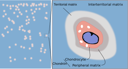
Cells that form the cartilage are called chondrocytes. They are located through the tissue in scattered small cavities known as lacunae. Chondrocytes are round to ellipsoid cells with many irregular microvillosities in the plasma membrane, and many bearing a cilium. Immature cartilagenous cells (chondroblasts) contain well-developed secretory organelles, such as rough endoplasmic reticulum and Golgi apparatus, for synthesizing and releasing collagen and elastic fibers. They also contain glycogen depots and lipid droplets in the cytoplasm. Chondrocytes are surrounded by a thin layer of pericellular extracellular matrix showing a distinct molecular composition. Chondrocyte and pericellular thin layer of extracellular matrix are together known as chondron or chondrome (Figure 1). Unlike osteocytes, chondrocytes are not connected by cytoplasmic processes. Extracellular matrix is synthesized by chondrocytes and chondroblasts, and can be removed by chondroblasts. Most chondrocytes are able to divide, but it is infrequent. For example, cells of the articular cartilage that divide are less than 1 % of the total chondrocyte population.

Cartilage is surrounded by a layer of connective tissue known as perichondrium, excepting a type of cartilage known as fibrous cartilage. Perichondrium has an external layer, called fibrous perichondrium, composed of fibrous connective tissue containing collagen fibers and fibroblasts, and an internal layer called chondrogenic perichondrium, where chondrogenic cells and chondroblasts are found. Chondrogenic cells differentiate into chondroblasts, and chondroblasts become chondrocytes. Chondroblasts synthesize most of the new extracellular matrix. During their differentiation, chondroblasts get surrounded by their own extracellular matrix and become chondrocytes. This growth is known as appositional growth. In young cartilage, however, chondrocytes can proliferate and contribute to the synthesis of extracellular matrix. This type of growth is referred as interstitial growth.
Three types of cartilage have been found in mammals: hyaline, elastic and fibrous cartilage. The variety of cartilages is wider in other vertebrates.
2. Hyaline cartilage
Hyaline cartilage is the most widely distributed type of cartilage through the animal body. It is usually associated with bones. During development, hyaline cartilage forms the skeleton of vertebrate embryos. In adults, it can be found in tracheal rings, bronchi, nose, larynx, articular surfaces, and in the joints between the sternum and ribs. As animal gets old, hyaline cartilage loses water content and necrotic areas may appear in the central parts of the cartilage. Hyaline cartilage can grow and be repaired, as long as perichondrium is preserved.

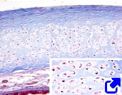
Hyaline cartilage is made up of mature cartilage, which account for most of the cartilage, and perichondrium, that covers the outer surface of the mature cartilage. The extracellular matrix of the hyaline cartilage shows uniform appearance. Type II collagen and proteoglycans are abundant, and other types of collagen are also present. Extracellular matrix is released by chondrocytes located in cavities known as lacunae. Chondrocytes are round to ovoid, and are usually found in couples or tetrads, known as isogenous groups (Figure 2). These groups are separated between each other by the so-called interterriotorial extracellular matrix. Type IV collagen and proteoglycans are abundant in the extracellular matrix near the isogenous groups (Figure 1), but type II collagen is scarce.

Perichondrium is a layer of dense connective tissue that covers the outer surface of the mature cartilage. The outer part of perichondrium is known as fibrous because it is composed of collagen fibers, some fibroblasts, and a net of blood vessels. The inner part of perichondrium is referred as chondrogenic because new chondrocytes arise from this layer and become part of the mature cartilage while they synthesized extracellular matrix.
Articular cartilage

Articular cartilage is a type of hyaline cartilage found in the synovial joints (they support frequent movements). Articular cartilage lacks perichondrium and grows from a population of progenitor cells found in the cartilage surface. Curiously, this peripheral population can also differentiate in bone, tendon and perimisium. The main function of articular cartilage is to resist the mechanical loads and provide a smooth and lubricated surface for reducing the effect of rubbing during body movements. Articular cartilage is made up of several layers (Figure 3). The outer one is in contact with the synovial liquid, it consists of non-mineralized extracellular matrix with long and crossed collagen fibers. The middle layer is thin and irregular with the extracellular matrix showing some mineralization. The inner layer is in contact with the bone tissue, and consists of mineralized extracellular matrix that is continuous with the extracellular matrix of the bone. While the outer layer is to minimize the effect of rubbing, the middle layer and, particularly, the inner layer, withstands the mechanical loads. Extracellular matrix, besides water, contains abundant type II collagen and proteoglycans, mainly aggrecan and chondroitin sulfate. The pericellular matrix of chondrocytes of the articular cartilage lacks type II collagen.

3. Elastic cartilage

Elastic cartilage (Figure 4) contains a high amount of elastic fibers so that it can be stretched while keeping its structural integrity. It is found in the outer ear, Eustachian ducts, epiglottis, and larynx. Elastic cartilage shows little extracellular matrix containing highly amount of branched elastic fibers that may account for 20 % of the dry weight and contribute to the mechanical properties of the tissue. Type II collagen is the most abundant collagen. Elastic cartilage does not arise from chondrogenic centers, but directly from mesenchymal tissue. The perichondrium is a sheath of very dense connective tissue lining the outer part of the cartilage. Isogenous groups, 2 to 4 chondrocytes, are not easily distinguished. Elastic cartilage does not become bone and it is not capable of self-repairing.

4. Fibrous cartilage

The fibrous cartilage is found in intervertebral discs, some joints, insertion of tendon into the epiphysis of bones (Figure 5), in the heart valves, and in the penis of some animals. It is usually surrounded by hyaline cartilage and shows mechanical properties between dense connective tissue and hyaline cartilage. Fibrocartilage cells are found more scattered than in the hyaline cartilage, but they are also distributed in rows, and sometimes it is difficult to distinguish chondrocytes from fibroblasts. The ultrastructural features of fibrous cartilage are similar to those of hyaline cartilage.

Four to seven types of collagen have been found in fibrous cartilage depending on where it is located. Type I collagen is the most abundant, accounting for about 80% to 90 % of total collagen. The high proportion of type I collagen (typical of tissues under strong mechanical tension) compared with type II collagen (abundant in tissues with heavy mechanical pressures) is a feature of fibrous cartilage. Collagen fibers are oriented toward the direction of the mechanical forces. There are not many elastic fibers and the major components of the ground substance of the extracellular matrix are proteoglycans, although they are less abundant than in the hyaline cartilage. The proportion of ground substance is lower than in other cartilages, which allows better visualize the collagen fibers. Fibrocartilage is less elastic than hyaline cartilage, but more than tendons. It is formed from precartilage, hyaline cartilage, and from fibrous tissue, depending on the body region.
-
Bibliography ↷
-
Bibliografía
Benjamin N, Evans EJ. 1990. Fibrocartilage. Journal of anatomy. 171: 1-15.
Fox AJS, Bedi A, Rodeo SA. 2009. The basic science of articular cartilage: structure, composition, and function. Sport health. 1: 461-468.
Salter DM. 1998. Cartilage. Current Orthopaedics, 12(4), 251-257.
Wilusz RE, Sanchez-Adams J, Guliak F. 2014. The structure and function of the pericellular matrix of articular cartilage. Matrix biology, 39, 25-32.
-
 Adipose