

Adipose is a specialized connective tissue that functions as the major storage site for lipids. It can be regarded as a rather unusual connective tissue since it has very little extracellular matrix. However, it is developed from mesenchymal cells derived from mesoderm during embryonic development, the same cells that give rise to the other connective tissues. Adipose tissue is present in mammals and some non mammal animal species. Adipocytes are the cells that form the adipose tissue, and they have the ability of synthesizing and storing large lipid droplets in the cytoplasm. Fat is a very suitable storage material because it contains approximately twice amount of energy than carbohydrates and proteins. These fat depots provide lipids that are used by other tissues to produce energy or just heat. Adipocytes are usually found in large and dense groups to form adipose tissue, although they can also be found scattered in the loose connective tissue. Besides adipose tissue, the second larger storage of lipids is the liver.
Adipose tissue is not just for lipid storage. It is also involved in the control of the body metabolism by releasing several hormones, cytokines, proteins, specific lipids, and mi-RNAs.
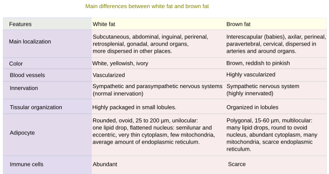
Two types of adipose tissue are found: white fat (or unilocular) with adipocytes containing a large lipid droplet, and brown fat (or multilocular) with adipocytes containing many small lipid droplets. The white color, some times yellowish, or brown of the fat is when the tissue is fresh. White and brown fat have particular features (Figure 1). Hay dos tipos de tejido adiposo: el formado por grasa blanca (o unilocular), cuyos adipocitos presentan una gran gota de lípidos, y el formado por grasa parda (o multilocular). El color blanco (a veces amarillento) o pardo se refiere al color de la grasa en su estado fresco. Ambos tipos de grasa tienen características particulares (Figura 1).
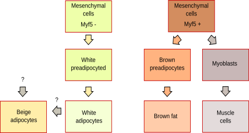
It is not well-known the differentiation paths of adipocytes from stem cells. However, both differentiate from mesenchymal cells but from different mesenchymal cell types. In fact, brown fat adipocytes share progenitor cells with muscle cells (Figure 2).
1. White fat
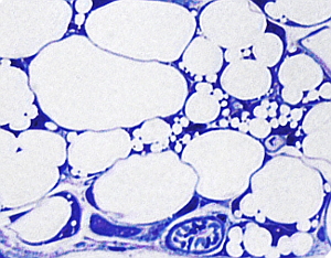
White adipose tissue (or unilocular adipose tissue) is the most abundant fatty tissue of mammals. Adipocytes form the white fat tissue. They are round and very large cells (more than 100 µm in diameter) containing one large lipid droplet that occupies most of the interior of the cell. That is why they are known as unilocular adipocytes. The nucleus and the remaining cytoplasm are found in a thin layer close to the plasma membrane (Figure 3). In well-fed animals, some adipocytes with more than one lipid droplets are observed among the typical white adipocytes (Figure 3). These adipocytes with more than one lipid droplet are not classified as multilocular or brown adipocytes (see below) but as white adipocytes showing an ongoing differentiation. During this differentiation process, adipocytes produce many small lipid droplets in the cytoplasm that will join in on large droplet in mature adipocytes. Thus, white fat adipocytes go through a multilocular period during differentiation. Much less common among white fat adipocytes are the beige adipocytes, probably derived from white fat adipocytes, but with the same features as brown adipocytes.

Unilocular adipocytes are separated between each other by very thin layers of loose connective tissue abundant reticular fibers released by the adipocytes themselves. Furthermore, each adipocyte is wrapped by a thin layer of extracellular matrix found very close to the plasma membrane. This sheath is called external lamina, and it is different from the surrounding connective tissue. The external lamina is similar to the epithelium basal lamina. In those parts of the body under mechanical stress, adipocytes are grouped in lobes separated by layers of connective tissue known as septa (Figure 4), which are more or less thick depending on the mechanical forces that they have to withstand. Each lobe usually contains large groups of adipocytes. Other cell types, such as mastocytes, macrophages, leukocytes, dispersed fibroblasts, and undifferentiated adipocytes, can also be found among the mature white adipocytes and in the septa.

Blood vessels and nerve fibers run through the connective tissue among the white adipocytes. Lymphoid nodes can be observed in the white fat of mesenteric areas. White fat is highly irrigated by blood vessels, as much as the muscle tissue. The endothelium of the capillaries is continuous. White fat receives two types of nerve inputs: one is coming from the sympathetic nervous system and the other from sensory axons coming from the dorsal spinal ganglia. Nerves do not contact adipocytes but end around the blood vessels.

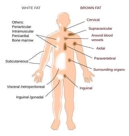
Although white fat tissue can be found in different locations of the body, it is mainly found in the subcutaneous and abdominal regions. Dermal and bone depots are also prominent. In humans, women and men have some common fat depots, but differences in the distribution are also found (Figure 5). There are some evidences suggesting that different fat depots of the body perform different functions, and it is the distribution of fat, more than the amount of fat, what may cause metabolic pathologies. For example, accumulation of fat in the visceral depot and the abdominal subcutaneous depot increases the risk of type II diabetes and cardiovascular diseases. However, gluteus and femoral depots may be protective fat. Internally, white fat is abundant in the mesenteries and in the intraperitoneus, and it is present, but less abundant, in the bone marrow and around inner organs. Subcutaneous depot, besides being an energy storage, works as an insulating layer against the cold. Fat depots in soles and in palms mainly perform a protective function against mechanical forces, while storage is less relevant. The dermal depot is different from subcutaneous depot, and they are physically separated. Dermal depot is involved in wound repairing, generation of hair follicles and termogenesis. In the bone marrow, there are two types of fat depots: constitutive and regulated. They are both engage with the bone physiology (mineralization and osteoclast activity) and are an important source of circulating adiponectin.
White adipose tissue is one of the few tissues that can increase and decrease its volume dramatically during the adult period. It is mainly a consequence of the increase in size of adipocytes (hypertrophy), as well as a recruitment of new adipocytes by proliferation of precursor cells (hyperplasia). Precursor cells are found in the connective tissue that surround adipocytes. In athletes, the white fat may be up to 2 to 3 % of the body weight, whereas an obese person may reach up to 60-70% of the body weight. Between 9 and 18 % of the body weight in men and between 14 and 28 % in women are considered as normal. It is obesity if the percentage of white fat is higher than 22 % in men, and 32 % in women. The hypertrophy and hyperplasia is different depending on the fat depot, even in the same depot between women and men.
White adipose tissue also performs an endocrine function by releasing some molecules called adipokynes that influence the insulin activity and the general homeostais of metabolism. For example, the leptin hormone controls the intake of food by acting in the central nervous system. There are other like resistin and adiponectin. In turn, white adipose tissue is regulated by other hormones like noradrenaline and glycocorticoids, which facilitate the release of fatty acids from adipocytes, and insulin that favors triacylglycerides storage. Adipocytes also store fat soluble vitamins.
In mice, it has been reported that in cold environments white fat adipocytes of inguinal fat depots may differentiate into cells with features similar to brown fat adipocytes. This happens by a transdifferentiation process, not by proliferation of undifferentiated progenitors. This multilocular adipocytes are known as brite or beige adipocytes. They can express the UCP1 protein that uncouples the ATP synthase in mitochondria, so that the proton gradient is used for heat production.
2. Brown fat
Brown fat is made up of adipocytes containing many small lipid droplets in the cytoplasm. Microscopy images show these cells with many holes inside because the standard histological processing removes lipids from the tissue. Therefore, the name multilocular adipocytes. Brown fat is abundant in hibernating animals, in fetuses, and in perinatal mammals. However, it is very scarce in adults. During development, brown adipose tissue appears earlier than white adipose tissue.
Brown fat adipocytes are smaller than white fat adipocytes, and their nucleus is rounded and located in central places of the cytoplasm. The brown color of fresh brown fat tissue is provided by the large number of mitochondria of adipocytes with contain abundant cytochrome oxidase. The dense vascularization also contribute to the brownish color. Brown fat adipocytes are characterized by expressing the UCP1 protein, which uncouples the electron transporters chain from the synthesis of ATP, so that the energy of generated proton gradient is used for heat production.
The appearance of the brown fat adipocytes may change under different conditions. For example, the adipocytes of well-fed animals kept in warm environments look like white fat adipocytes. It the animal is placed in a colder environment, brown fat adipocytes return to the multilocular morphology and increase in number.
In humans, brown adipose tissue is widespread in two wide regions:
Visceral: perivascular (arteries: aorta, carotid, brachiocephalic, epicardial coronary, internal mammary, intercostal ; veins: cardiac and intercostal ), surrounding hollow organs (heart, trachea, large bronchi, mesocolom, omentum principal), and around solid organs (paravertebral torax, pancreas, kidney, liver hilus).
Subcutaneous: anterior muscles of the neck, supraclavicular fossa, under the clavicle, axilla, anterior abdominal wall, inguinal fossa.

Brown adipose tissue is divided in large and small lobes separated by connective tissue. Blood vessels, more abundant than in the white fat tissue, and nerves run through this connective tissue. Brown fat is innervated by the sympathetic nervous system, that after releasing noradrenaline leads to brown adipocytes to degrade lipids and generate heat. Unlike in white fat tissue, terminals of the sympathetic axons directly stimulate brown fat adipocytes.
-
Bibliografía ↷
-
Bibliografía
Frühbeck G, Sesma P, Burrell MA. 2009. PRDM16: the interconvertible adipo-myocyte switch. Trends in cell biology. 19: 141-146.
Gesta S, Tseng Y, Kahn CR. 2007. Developmental origin of fat: tracking obesity to its source. Cell. 131: 242-256.
Hausman DB, DiGirolamo M, Bartness TJ, Hausman GJ, Martin RJ. 2001. The biology of white adipocyte proliferation. Obesity review. 2: 239-254.
Rosenwall M, Wolfrum C. 2014. The origin and definition of brite versus white and classical brown adipocytes. Adipocyte. 3: 4-9.
Sanchez-Gurmaches J, Hung C-M, Guertin DA. 2016. Emerging complexities in adipocyte origins and identity. Trends in cell biology. 26:5
Sacks H, Symonds ME. 2013. Anatomical locations of human brown ddipose tissue. Functional relevance and implications in obesity and type 2 diabetes. Diabetes. 62:1783–1790
Saely CH, Geiger K, Drexel H. 2010. Brown versus white adipose tissue: a mini-review. Gerontology. 58: 15-23.
-
 Connective proper