Animal tissues
Connective
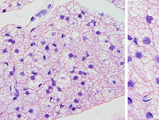
BROWN ADIPOSE

Species: mouse (Mus musculus; mammal).
Technique: haematoxylin-eosin, 8 μm thick section, paraffin embedding.
Brown fat tissue is made up of multilocular adipocytes, which contain many lipid droplets in the cytoplasm (white fat adipocytes have just one lipid drop, that is, they are unilocular). The nucleus is rounded and the cytoplasm is pink when stained with eosin. Brown fat tissue is highly irrigated by blood vessels, that together with the large amount of mitochondria in brown adipocytes, make this tissue to be brownish. That it is why the name brown adipose tissue. Adipocytes are arranged in lobes separated by connective tissue. The function of the brown adipose tissue is not to store energy for metabolism, but the fat is consumed to generate heat. In most mammals, brown adipose tissue is abundant during the perinatal period, and progressively dissapears during development. In adults is restricted to a few places of the body.