Animal tissues
Connective
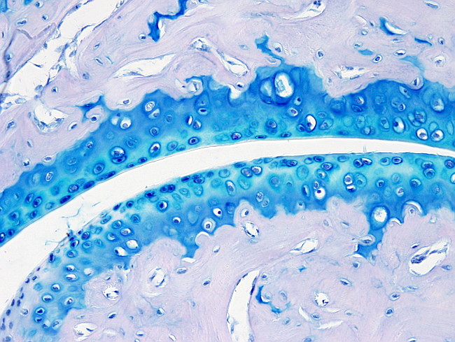
ARTICULAR CARTILAGE

Species: mouse (Mus musculus; mammal).
Technique: paraffin sections stained with hematoxylin-tolouidin blue.
The articular cartilage is found in the synovial joints. It lacks perichondrium and its main function is to absorb the mechanical pressures and provides a surface for friction between bones during the body movements. Like other hyaline cartilages, the articular cartilage is not irrigated by blood vessels, lymphatic vessels, and it is not innervated by the nervous system. In addition, it has low regenerative capacity. The thickness of the articular cartilage is variable depending on the species, but it is usually about 1 to 2 mm.
The extracellular matrix of the articular cartilage contains collagen fibers, glycosamincoglycans and glycoproteins. These molecules are responsible for the mechanical properties of the tissue and help to increase hydration, which is essential for the articular cartilage functions. The proportion of water decrease from the superficial region (80%) to the deep region (65%)(see below). The collagen is the most abundant organic molecule, particularly the type II collagen, but the I, IV, V, VI, IX y XI collagen types are also present, which stabilize the type II collagen. Proteoglycans are up to 10 to 15 % of the dry weight of the articular cartilage, being the aggrecan the most abundant. In general,chondrocytes are up to 2 % ot the total articular cartilage volume.
Several regions are distinguished in the articular cartilage. The superficial region is the outermost part that physically contacts with the superficial region of the articular cartilage of opposite bone in the joint. It is about 10 % to 20 % of the total thickness of the articular cartilage. Collagen types II and IX are abundant in this region. Chondrocytes, the mature cartilage cells, are flattened. The superficial region is in contact with the synovial liquid of the joint. The middle region follows the superficial region, encompassing about 40 to 60 % of the cartilage thickness. It contains many proteoglycans and thick collagen fibers and shows less chondrocyte density. The middle region is the main responsible for withstanding mechanical pressures. The deep region is up to 30 % of the cartilage thickness. It is the layer closer to the bone. The collagen fibers are the thickest and are arranged perpendicularly to the articular surface. Many proteoglycans are also present. The deep region is the strongest layer. Chondrocytes are usually lined up into columns perpendicular to the cartilage surface. Between the deep region and the bone, there is region of calcified cartilage that anchors the articular cartilage to the bone. Entre la zona profunda cartilaginosa y el hueso está situada la zona de cartílago calcificado, la cual ancla el cartílago al hueso.