Animal tissues
Connective proper
DENSE REGULAR

Species: mouse (Mus musculus; mammal).
Technique: 8 µm thick paraffin sections stained with hematoxylin and eosin.
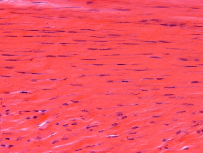
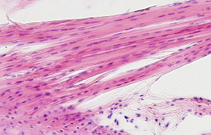
In this image of a tendon, it can be observed that most part of the tissue is extracellular matrix (reddish color provided by eosin). Fibroblasts (dark color provided by hematoxylin) show flattened nucleus, clearly observed in the upper part of the image. There are very few clear spaces in the tissue, indicating that the extracellular matrix is highly packaged. That is why is called dense connective tissue. In addition, collagen fibers are organized parallel to each other. Therefore the name regular. For instance, in the upper part of the image, collagen fibers are parallel to each other and to the section surface, and fibroblasts are found organized as rows, adapted to the fiber disposition.
In tendons, the extracellular matrix is composed of collagen fibers (65-80% of dry weight), mainly type I collagen, and elastic fibers (1-2 % of dry weight), both embedded in ground substance which contains proteoglycans and water. In humans, microfibrils are 60 to 175 nm in thickness. Connecting bridges are established between microfibrils that make strong collagen fibers. It is of notice that collagen fibers can be stretched only 4 to 5 % of their length. Fibroblasts in tendons are known as tenoblasts or tenocytes (depending on their differentiation stage), and their morphology is adapted to the orientation of collagen fibers. They release collagen and elastin, so they show abundant endoplasmic reticulum and many ribosomes.
Tendons are anchored to the fibrous part of the periostium by one end, although some collagen fibers may be seen entering the bone matrix. Cartilage cells can be observed near the bone-tendon junction. Tendons are repaired easily, and they can be used for transplantation.
Images


 The image is from a tendon.