Animal organs. Cardiovascular.
ARTERIOLE

Species: rat. (Ratus norvegicus; mammal).
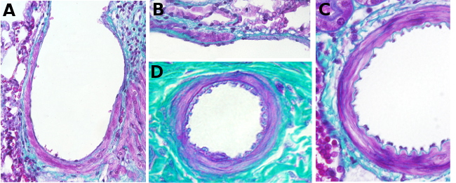
Technique: paraffin embedding, staining with Masson trichome.
Arteries, the blood vessels conducting the blood from the heart to the organs, show different diameters. The larger arteries are known as elastic arteries and the middle size arteries as muscular arteries. Arterioles have a smaller diameter, less than 100 µm, and they progressively decrease in size until they become capillaries.

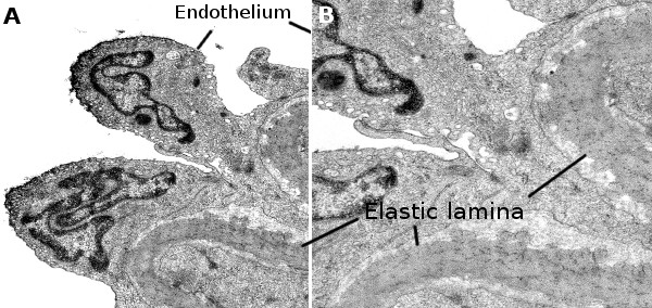
Like other arteries with larger diameter, arterioles are made up of three layers or tunica. The inner tunica, in contact with the blood, is the tunica intima, which is composed of endothelium and a layer of connective tissue. The tunica media is located more externally and is mainly formed by smooth muscle cells, oriented parallel to the longitudinal axis of the arteriole, and by fibroelastic connective tissue. Unlike larger arteries, the arteriole tunica media is poorly developed and has less than three layers of smooth muscle cells. By definition, when more of two layers of smooth muscle cells are found in the tunica media the name of the blood vessel changes to small artery, and to muscular artery if it is more than 8-10 layers. The tunica adventitia is the most outer layer and it is mostly fibroelastic connective tissue. Sometimes, the tunica adventitia is difficult to be distinguished.
Arterioles distribute the blood to the capillary nets and are in charge of the blood pressure (arterial tension). By cell contraction, the smooth muscle cells of the tunica media decrease the blood flow to capillaries. In the interface between arteriole and capillary, there is a light increase in the amount of smooth muscle cells forming the so called precapillary sphincter. Arterioles are able to distribute the blood where it is needed by regulating the blood flow. For example, to the intestine for digestion, to muscles for exercise, or to the brain for a higher neuronal activity.
In the hypertension pathology, the diameter of the arterioles and small arteries is smaller because the vessel wall increase in size. This may be a consequence of the deposition of lipids among the smooth muscle cells or by the increase in muscle cells number. Both pathological processes can be found at the same time.
