Exocytosis is the fusion of vesicles with the plasma membrane. Vesicles for exocytosis are mostly shipped from the trans domain of the Golgi apparatus, are moved to the cell periphery and fuse with the plasma membrane. Vesicles can also be shipped by other organelles, like endosomes (see below).
1. Types of exocytosis
There are two types of exocytosis: constitutive and regulated (Figure 1). Constitutive exocytosis is present in almost every cell and carries molecules needed by the plasma membrane and the extracellular matrix. It is the default exocytic pathway, a continuous trafficking where the amount of vesicles depends on the physiological state of the cell. Regulated exocytosis is present in cells specialized in secretion, such as endocrine cells, neurons, intestine epithelial cells, and glandular cells. Through regulated exocytosis, molecules are released into the intestine lumen for digestion, or, like hormones, they are released into the extracellular matrix to modulate the physiology of other cells located either close or quite far in the body by traveling through the circulatory system. Vesicles of regulated exocytosis fuse with the cell membrane after a signal, which is usually an increase in the cytosolic calcium concentration. So, they do not fuse spontaneously. Furthermore, ATP and GTP are needed as energy sources.

2. Selecting cargoes in the Golgi apparatus
Constitutive and regulated exocytosis pack different types of molecules. The TGN must be able to sort both types of cargoes in different vesicles. Constitutive vesicles enclose those molecules without a specific signal. However, not all these molecules are transported in the same way. For instance, there are vesicle departing from the TGN with their membranes enriched in sphingomyelin. These vesicles may bud from domains of the TGN membranes having many sphingomyelins. Lipase lipoprotein is a soluble protein abundantly packaged into these vesicles because it has a molecular domain that bind heparan sulfate. Heparan sulfate is part of the proteglycan syndecan, which binds to sphyngomyelin enriched domains. In this way the lipase lipoprotein associates to syndencan, which in turn binds to sphyngomyelin, and both are packaged in the same type of vesicles.
How are cargoes selected for regulated exocytosis? It has been suggested that molecules for regulated exocytosis form large molecular aggregates, which include precursors and enzymes. Many molecules are incorporated in regulated exocytosis vesicles as inactive forms, such as pro-neuropeptides. Enzymes process precursors inside the vesicle. Furthermore, in the TGN, there are also molecules targeted to endosomes, fetched by clathrin coated vesicles, and others targeted to the endoplasmic reticulum, which are included into COP-I coated vesicles.
3. Regulated secretion
Regulated exocytosis vesicles are released from the Golgi apparatus and remain in the cytoplasm. After the cell receives a signal, vesicles are moved to specific areas of the cell periphery and fuse with the plasma membrane. So, it is a regulated process not only temporally but also spatially. Neurons are a good example of regulated exocytosis. In these cells, some vesicles are formed around the nucleus, in the soma, and are transferred to the presynaptic terminal, which can be several centimeters away from the soma. Other polarized cells are the enterocytes of the intestine epithelium, which have an apical domain facing the lumen of the intestine and a basolateral domain. It would be a catastrophe if vesicles loaded with digestive enzymes are released into the basolateral domain because the surrounding tissue would be digested. Regulated exocytosis vesicles are moved to the correct domain of the plasma membrane by microtubules and actin filaments of the cytoskeleton, helped by motor proteins.
Exocytosis involves the fusion of a vesicle with the plasma membrane, and then the vesicle membrane molecules become part of the plasma membrane. However, electron microscopy images suggest another exocytosis mechanism, which has been named "kiss and run" (Figure 2). This model proposes that vesicles do not end as part of the plasma membrane because the fusion process is transient. There is an initial fusion between the vesicle and the plasma membrane that affects a small area of both membranes. A small pore is opened allowing the communication between the interior of the vesicle and the extracellular space, through which soluble molecules are released by diffusion. The pore is transient and gets closed after a while, vesicle and plasma membranes are sealed again, and the vesicle is free and empty in the cytosol. The kiss and run mechanism is thought to occur in many cell types, particularly in synaptic terminals of neurons and in chromaffin cells. It has also been observed that vesicles can be fused between each other before the fusion with the plasma membrane (Figure 2). This mechanism is known as compound exocytosis because the content of several vesicles is mixed before it is exocytosed.

4. Other sources of vesicles
Not every vesicle that gets fused with the plasma membrane is shipped by the Golgi apparatus. Early endosomes are organelles targeted by endocytic vesicles (vesicles formed in the plasma membrane)(Figure 3). After the fusion with the endosome, some of the endocytosis vesicle content, particularly membrane proteins and lipids, is sent back to the plasma membrane by vesicles budding from the endosome. Another example of exocytosis independent of the Golgi apparatus is found in the synaptic terminals of the nervous system (Figure 4). Synapses are usually far away from the Golgi apparatus, which is found in the neuronal soma. Neuronal communication cannot rely on a long and slow transport of neurotransmitters from the soma to the synaptic terminal, because the communication between neurons would be too slow and inefficient. Thus, there is a local mechanism for exocytosis in the synaptic terminals. It starts with vesicle formation by endocytosis in the plasma membrane, near the synaptic density (where neurotransmitter are going to be released). Once vesicles are in the cytoplasm of the synaptic terminal, they are loaded with neurotransmitters that cross the membrane vesicle through transporters found in the vesicle membrane. Once loaded, vesicles are transferred near the presynaptic density, where they are ready to fuse with the plasma membrane (exocytosis) after the action potential arrives. These vesicles release small neurotransmitters. In this way, there is a continuous process of vesicle formation, loading and exocytosis (release of neurotransmitters) in the synaptic terminal, so it is much more efficient.


5. Organelle fusion
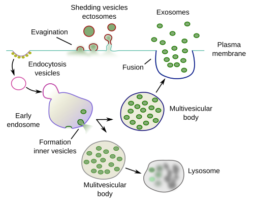
Multivesicular bodies, which are endosomes containing vesicles, can occasionally get fused with the cell membrane and release their vesicles into the extracellular space (Figure 5). These vesicles are known as exosomes. This exocytosis mechanism was described in the 1980s, and the name exosomes was given to these vesicles. It was first observed during the maturation of reticulocytes to erythrocytes.

Under some circumstances, other organelles can get fused with the plasma membrane. For example, when plasma membrane is damaged, and there is a hole, cells has to seal that membrane gap. Lysosomes, endosomes and endoplasmic reticulum can get fused with the plasma membrane to repair membranes by adding and sealing the breakages. Similarly, cells need to increase enormously the surface during phagocytosis to engulf a particle. The surface of the plasma membrane is increases by fusion of endoplasmic reticulum tubules and cisterns, as well as the other organelles just mentioned.
-
Bibliography ↷
 Golgi apparatus