Animal tissues
Glandular epithelium
PANCREAS

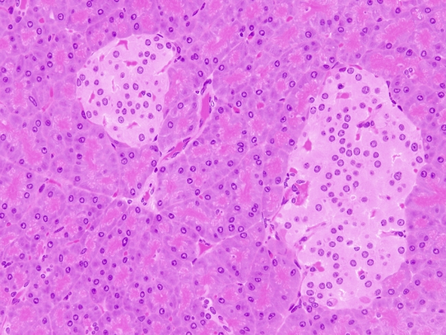
Species: mouse (Mus musculus; mammal).
Technique: 8 µm paraffin section stained with haematoxylin-eosin.
The pancreas consists in an exocrine component, the pancreatic acini, and an endocrine part composed of the Langerhans islets. That is why it is named as amphicrine gland The major part of pancreas corresponds to the exocrine part (98-99 % in humans). The serous exocrine cells show a rounded nucleus and dark cytoplasm. The cytoplasm is more clear in the apical part because of the higher density of secretory zymogen granules containing digestive enzymes (zymogens are inactive enzymes or proenzyme). Abundant rough endoplasmic reticulum is localized in the basal cytoplasm of serous cells, where enzyme precursors are synthesized, that gives a darker color. Within serous acini, there are weaker stained cells which are named as centroacinar cells. They form ducts to collect the secretory substances released by serous cells. These acinar ducts join together and form the interlobular ducts, which in turn flow into the hepatic ducts, and finally release the pancreatic secretory products into the gut. There are no myoepithelial cells around the pancreatic acini.
The endocrine part of the pancreas is made up of cell groups which are known as islets of Langerhans. They are distributed among the exocrine acini. The islets of Langerhans are highly irrigated by blood vessels that uptake and distributed the hormones released by the Langerhans islet cells. Although it is difficult to discern using common staining techniques, there are several types of cells in the islets of Langerhans: α cells (release glucagon), β cells (release insulin), gamma cells (release somatostatin) and F cells (release pancreatic polypeptide).

 This image is from pancreas.