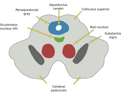
The mesencephalon is found between the rhombencephalon and the diencephalon (Figure 1). It is also known as the midbrain. In humans, it is 2.5 cm in length. Despite its small size, many essential functions are performed by the mesencephalon, such as vigil, response to pain, motor coordination, control of eye movement, and vision and sound processing. In addition, many axonal tracts travel through the mesencephalon, both sensory bundles toward the thalamus and descendant bundles from the cerebral cortex.

Along the dorsoventral axis, the mesencephalon is anatomically divided into the alar plate (dorsal) and basal plate (ventral). The four culliculi (corpora cuadrigemina) are found in the most dorsal part of the alar plate and the tegmentum in the basal plate. In humans, there are prominent columnar structures in the ventral mesencephalon, known as cerebral peduncles, which contain the descending cortical projections (pyramidal pathway).
At the mesecephalon roof (most dorsal alar plate), the corpora cuadrigemina consists of four small protrusions known as colliculi, two superior (rostral; Figure 2), and two inferiors (caudal). The superior colliculi are involved with vision and eye reflexes. In those animals lacking visual cortex, the superior colliculi are the main responsible for visual information processing. They receive information from the retina, cerebral cortex, as well as from the rhombencephalon and spinal cord. Damages in the superior colliculi lead to the inability to follow the movement of an object with the eyes. The inferior colliculi, or torus semicircularis, are involved in the auditory information. The neurons of the inferior colliculi are arranged in layers. Damages in both inferior colliculi leads to deafness and in one collicilus does no allow to localize the source of the sounds in the 3D space.

The tegmentum is found in the basal plate. It contains the periaqueductal gray, which is a group of nuclei around the mesenphalic aqueduct that extends rostrally toward the diencephalon and caudally toward the rhombencephalon. These nuclei are organized in four longitudinal columns and perform several functions, but generally, this region is related to pain processing.
The reticular formation is also found in the tegmentum. It is an elongated structure in the ventral midline, composed of several neuronal groups. There are rostral and caudal regions of the reticular formation specialized in different functions.
The red nucleus is a group of neurons found in the rostral and central tegmentum. The red color is because of dense blood vessel vascularization (more iron). It is involved in motor control. It receives inputs from the motor cortex and cerebellum and sends processed information to the motoneurons of the spinal cord.
The substantia nigra is found in the ventral tegmentum, just above the peduncles. The dark color is a consequence of the high content of melanin. In adult mammals, about 75 % of the dopaminergic neurons localize in the mesencephalon, more abundant in the pars compacta of the substantia nigra (SNc), which send projections to the dorsal striatum (subpallium; nigro-striatal pathway) and are involved in voluntary movements. These dopaminergic neurons are lost during Parkinson's disease. The other two large dopaminergic regions are the ventral tegmental area (VTA) and the retrorubral field (RRF), which send information to the ventral striatum and the entorhinal cortex (mesocorticolimbic pathway). These connections are related to emotional and reward behavior. Pathologies such as schizophrenia, addiction, and depression are influenced by these circuits.
The trochlear nucleus is found ventral to the periaqueductal gray and innervates the ocular muscles. The trochlear nerve (IV) crosses the mesecephalic midline and exits the encephalon through the dorsal region. These two features are unique when compared with the other cranial nerves.
The oculomotor nucleus is located in the mesencephalic tegmentum, under the rostral culliculi, and ventral to the periaqueductal gray. Its axons form the oculomotor nerve (III). In most vertebrates, the oculomotor nerve innervates four extraocular muscles (three rectus and one obliquous), the elevator of the superior eyelid, and the intrinsic eye muscles (ciliary muscles and pupil).
-
Bibliography ↷
-
Bibliography
Puelles L, Martínez S, Martínez de la Torre M. 2008. Neuroanatomía. Editorial Médica Panamericana S.A. ISBN: 978-84-7903-453-5.
-
 Rhombencephalon