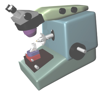
Very thin sections, known as ultrathin sections, are needed for studying the ultrastructural features of cells and tissues. These sections are usually about 40 to 80 nm in thickness and are studied with the transmission electron microscope. Ultrathin sections can be obtained from samples embedded in a very hard compound, like epoxy-type resins. After polymerization, resin blocks are sectioned with a special microtome, the ultramicrotome (Figure 1). It is a very sophisticated microtome that can push the sample toward the blade in very short distance intervals (nanometers). There is another apparatus, known as ultracryotome, that can get ultrathin sections from frozen tissue. It is used when some molecular features we are interested in are altered during the embedding process.

The ultramicrotome is designed for cutting by a mechanism similar to the paraffin microtome, but it is much more sophisticated and precise. So precise that it is possible to change the section thickness with differences of several nanometers. Currently, it is a mechanical device, but in older ultramicrotomes the advance of the sample over the blade was by heating the arm bearing the sample holder. By regulating the heat intensity, the arm grew in length by dilatation and so does the section thickness. In the current microtomes, the sample holder has a complete built-in system for orientating the surface of the sample with respect to the blade. Since sections are extremely thin, any vibration or temperature change may alter the thickness between successive sections. Thus, the ultramicrotome should be placed in a room free as possible of vibrations and with a regulated and constant temperature. Ultramicrote are usually held in a special table with a mechanism to absorb vibrations. There is also an external control panel for setting the sectioning process, including start and stop, thickness, cutting speed; cutting window, lights and other parameters.
Before the first section is obtained, as happened to paraffin embedding, a truncated pyramid has to be made in the resin block. This trimming process can be carefully performed with a sharp blade. However, there is a dedicated apparatus for this purpose that contains a milling system and a stereoscope. The trimming of the resin block must be very precise because the cutting surface needs to be very small, usually about 0.5 mm. The pyramid body with the trapezoidal surface is similar to those described for paraffin embedding, a larger cutting edge and a smaller parallel edge for getting long and straight rows of sections. Before obtaining ultrathin sections, it is common to get a thin section (0.5 to 1 µm in thickness) that can be visualized at light microscopy, so that a complete image of the tissue can be observed. This is a good practice to give context to the electron microscopy pictures or for proper orientation in the ultrathin section when observing with the electron microscope.
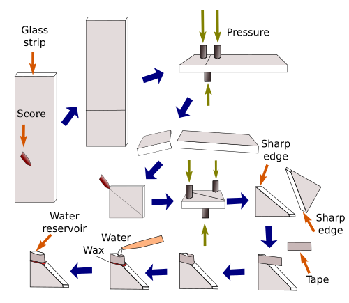
Ultramicrotome knives have very sharp cutting edges, needed to get ultrathin sections. They are usually made of special glass and are crafted from glass strips, by making streaks and pressures at precise points that break the glass strip in squares and then in knives (Figure 2). However, the higher quality of ultrathin sections is obtained with diamond knives, but they are much more expensive. Although the current ultramicrotomes are well-designed to get homogeneous ultrathin sections, it is common to get sections with slightly different thickness. How thick is an ultrathin section can be estimated by the color that the section reflexes from its surface. This color can be gray (less than 60 nm), silver (60 to 90 nm), pale gold (90 to 120 nm), dark gold (120 to 150 nm), and purple (150 to 190 nm).

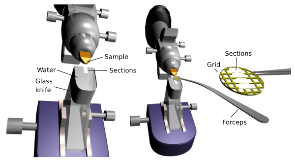
Ultrathin sections remain on the water surface of the knife water container (Figure 3). They are collected on cooper or nickel grids. The grids are rounded and thin, and they have a mesh that can be more or less dense, for small or large sections. The windows, spaces without bars, range from a hundred to several hundreds of µm2. These grids offer the best image quality because electrons only need to cross the section before hitting the visualization screen. However, the regions of the section overlapping the bars cannot be observed. There are other "grids" that have only one large window where the whole section can fit. Of course, a supporting film, made up of resin known as formvar, must cover the window. This film is very thin, lets the electrons going through and sustains the section.

 Cryotome