Vascular plants, unlike non-vascular plants, have specialized tissues for transporting water and both inorganic and organic substances. These tissues are known as vascular tissues, which include the xylem and phloem. The xylem is specialized in transporting water, inorganic compounds, and some organic substances from roots to other plant organs, while the phloem mostly transports organic substances synthesized in plant body organs, such as leaves and storage tissues, toward the rest of the plant.
1. Introduction
Physiologically, plants need vascular tissues to increase their size by distributing water and organic substances to feed the cells. These tissues also have a mechanical role in supporting the aerial parts and giving consistency to the underground organs, acting as a skeleton. Another function of vascular tissues is to allow communication between distant organs of the plant body by transporting meaningful molecules, like some phytohormones.
During the primary growth of the plant, the primary xylem and phloem are formed. The protoxylem and protophloem are the first functional vascular tissues of the plant that arise from the procambium meristem. It happens during embryonic development and at the tips of stems and roots during later development. The metaxylema and metaphloem differentiate from the procambium meristem too. They replace the protoxylem and protophloem as functional vascular tissues as the organs mature. If secondary growth takes place in the plant, the secondary xylem and secondary phloem are formed from the vascular cambium meristem, while the metaphloem and metaxylem become nonfunctional. Xylem and phloem, both primary and secondary, are always physically close to each other in all organs of the plant because they differentiate from the same meristem, either the procambium or the vascular cambium. These tissues are composed of several types of cells, some of which may provides some phylogenetic clues. The organization of vascular tissue in stems and roots is different.
The set of vascular tissues of a shoot or a root is known as the stele, and, depending on the organization, there are different types of steles. For instance, when the vascular tissues form a solid cylinder, the stele is known as the protostele, but if they form a hollow cylinder with parenchyma inside, the stele is known as the siphonostele (Figure 1).

2. Xylem
The xylem is responsible for transporting and distributing water and mineral salts, which mainly come from the root, to the plant body. It also transports some organic and signaling molecules. Furthermore, it is the main tissue for mechanical support of plant organs, particularly during secondary growth. The wood of trees and some plants is largely xylem.
Xylem consists of four cell types: a) vessel elements and b) tracheids are the conducting cells, also known as tracheary elements; c) parenchyma cells work as storing and communication cells; and d) sclerenchyma and sclereids are supporting cells.

Tracheary elements (a and b) are cells containing lignin in their thick and hard secondary cell walls. These cells lose their cytoplasmic content during differentiation. The secondary cell wall of the tracheary cells forms different thickenings depending on the cell type and developmental stage of the organ. Thus, vessel elements and tracheids are identified at light microscopy by the morphology of these thickenings, which can be annular, helical, reticulated, or dotted structures.
Vessel elements (a) are cells with a larger diameter and more flat ends when compared to tracheids (Figures 2, 3, and 4). They are connected longitudinally to one another to form long tubes, also known as vessels. Water is conducted through the symplastic pathway, that is, within the cells. The water crosses from one cell to the next through the perforation plates, which are the specialized perforated transverse cell walls at both ends of each cell. Furthermore, water can cross the lateral walls of the vessels through pits and pass to the lateral adjoining cells of the xylem. Vessel elements are the main conducting cells of the xylem of angiosperms.
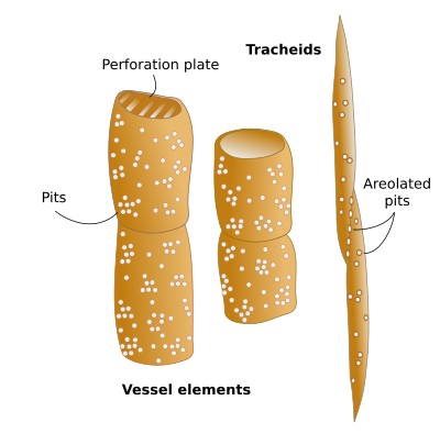
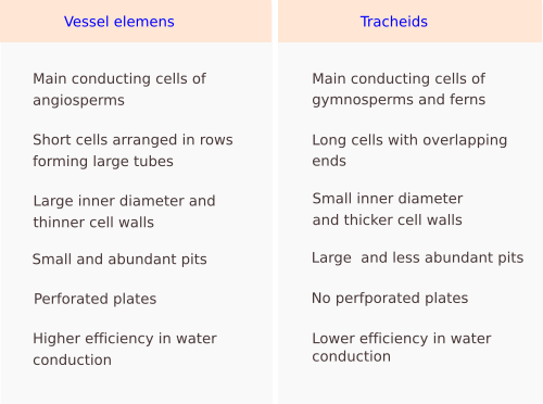
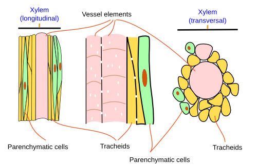
Tracheids (b) are the second conducting cell type of the vascular plants. It is the only tracheary element in pteridophytes and gymnosperms. Angiosperms have both tracheids and vessel elements. Tracheids are elongated, fusiform, thin cells. Water is conducted intracellularly and crosses from one cell to the next by symplastic transport through areolate pits found at the walls of both ends of each cell, which overlap with those of the previous and following cells (Figure 2). Water is also laterally transported through areolate pits located at the lateral cell walls. Because tracheids do not have perforation plates, they have a lower capacity for water conduction than vessel elements. Furthermore, tracheids have thicker cell walls and a smaller inner volume than vessel elements. Conifers show tracheids with very large and round areolate pits and an inner structure known as the torus, which is an oval thickening of the cell wall. The torus can regulate the flow of water through the pit.
Parenchyma cells (c) are organized in the conducting tissues in two ways: radially or axially. Radial parenchyma cells form rows, or rays, perpendicular to the surface of the organ, while axial parenchyma cell form groups, or rows, arranged longitudinally in the xylem, being more abundant in the secondary xylem (see below). Radial parenchyma cells are elongated, parallel to the axis of the ray, and connected to one another by numerous plasmodesmata that allow the transport of substances. In conifers, the rays are uniseriate or biseriate, i.e., consisting of one or two rows of cells, whereas in angiosperms, they are usually multiseriate with many rows of cells, sometimes with different types of cells. The rays of the xylem are continuous with the rays of the phloem. This is so because one single cell of the vascular cambium can differentiate into radial parenchyma cells of either the xylem or phloem.
Parenchyma cells perform multiple functions, like storing carbohydrates (starch), water, or nitrogen, and facilitating communication between xylem and phloem.
Sclerenchyma fibers and sclereids (d) provide support and protection.
The primary xylem is the first type of xylem developed during the formation of a plant organ. It is protoxylem first, and then metaxylem. The protoxylem is developed from the procambium meristem during organ growth. The protoxylem matures completely and disappears later due to the compression of mechanical forces that are produced by the growth of the organ. The secondary wall of conducting cells of the protoxylem (vessel elements and tracheids) initially shows annular thickenings that later become helical. The metaxylem appears after the protoxylem, when the organ enlarges and matures once the growth begins to slow. It also arises from the procambium. Their cells show larger diameters than those of the protoxylem, and the cell walls of the conducting cells have reticulated thickenings first and perforated thickenings later. The metaxylem is the mature xylem in those organs that don't go through secondary growth.

The secondary xylem is produced from the vascular cambium meristem in those organs with secondary growth. Together with secondary phloem, it is the mature conducting tissue in plants with secondary growth.
3. Phloem
The phloem, also known as sieve tissue or bast, consists of living cells. Its main role is transporting and distributing organic molecules synthesized by photosynthesis or mobilized from storing tissues, as well as signaling molecules like hormones.
Phloem is made up of more cell types than xylem. There are conducting and non-conducting cells. Conducting cells are the sieve cells (a) and sieve tubes (b) (Figures 5, 6 and 7), Both cell types are living cells, without nucleus, and their primary cell wall is thickened with callose deposits. Non conducting cells are parenchyma cells that include the abundant companion cells (c). There are also supporting cells (d) associated with the phloem, such as sclerenchyma fibers and sclereids.
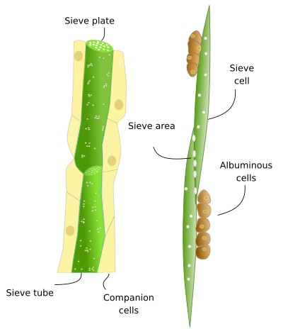
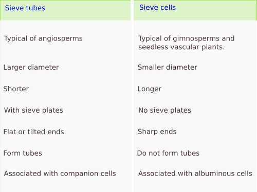

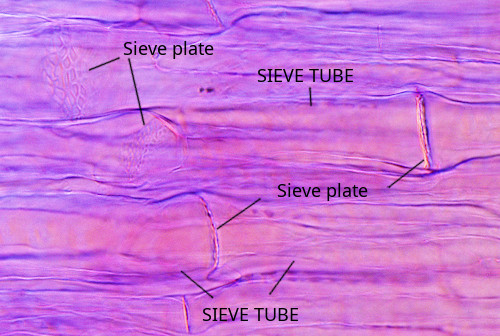
Sieve tubes (a) (Figure 8) are the typical conducting cells of angiosperms. They are formed of cells lined up into longitudinal rows that communicate with each other through sieve plates located at both ends (transverse) of each cell. Sieve plates have large-size pores that allow the direct connection between adjoining cytoplasms. In addition, there are sieve areas in the side walls that are discontinuities of the primary cell wall for communication with adjacent sieve tubes and with parenchyma companion cells. Sieve tubes are the main conductive element in angiosperms.
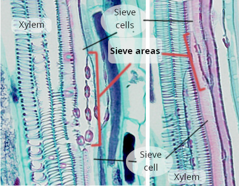
Sieve cells (b) (Figure 9) are typical of gymnosperms and pteridophytes. They are long cells with pointed ends, communicating with each other laterally by primary pore fields forming the sieve areas. They don't have sieve plates. Functionally and morphologically, sieve cells are associated with a type of specialized parenchymal cell called Strassburger's cells (albuminous cells). Sieve cells are the only conductive element in gymnosperms and pteridophytes.
Parenchyma cells (c) are components of the phloem. Companion cells are parenchyma cells strongly associated with the sieve tubes. They maintain the metabolism of sieve tubes since sieve tubes lack nuclei and show a reduced cytoplasm. Companion cells contain a large nucleus and a cytoplasm with abundant organelles, which means a high metabolic rate. However, they do not store starch. Companion cells and sieve tubes are differentiated from the same meristematic cells. Only angiosperms show companion cells. The cells associated with the conducting cells (sieve cells) in gymnosperms are known as Strasburger cells (albuminous cells), with similar functions to the companion cells.
Other types of parenchyma cells work as storage for substances transported by the phloem itself. In some species, there are cells in the phloem specialized for secretion. The interaction between parenchyma cells and conducting cells is strong, and when conducting cells die, parenchyma cells die too. In the primary phloem, parenchyma cells are elongated and vertically arranged, whereas in the secondary phloem there is an axial parenchyma, with elongated and vertically organized cells and a radial parenchyma with isodiametric cells.
Sclerenchyma fibers and sclereids (d) are also found in the phloem, with supporting and protection roles.
The primary phloem is the first phloem to be functional in developing organs. The first type of primary phloem to appear is the protophloem, which is later replaced by the metaphloem. Both the protophloem and metaphloem are formed from the procambium meristem. In angiosperms, protophloem contains non-well-developed sieve tubes and cells, while in gymnosperms and ferns, it shows poorly-developed sieve cells. Companion cells are rare or absent. Metaphloem replaces protophloem during development, usually when the organ stops growing. The metaphloem contains sieve tubes and sieve cells that are thicker and longer than in the protophloem. In angiosperms, it always contains companion cells. The sieve tubes have sieve plates. Metaphloem is the functional conducting tissue in plants with primary growth.
The secondary phloem arises from the vascular cambium meristem in plants with secondary growth. The conducting cells are well-developed, as are the companion cells, and both axial and radial parenchyma are present. Unlike in xylem, secondary phloem cells do not synthesize secondary cell walls, and therefore they are living cells. However, the cytoplasm of sieve elements may lack nuclei, microtubules, and ribosomes, and the border between the vacuole and the rest of the cytoplasm is not easily observed.
-
Bibliography ↷
-
Lalonde S, Francesch VR, Frommer WB. 2001. Commpanion cells. In: eLS. John Wiley & Sons Ltd, Chichester. http://www.els.net [doi: 10.1038/npg.els.0002087]
Furuta KM, Hellmann E, Helariutta Y. 2014. Molecular control of cell specification and cell differentiation during procambial development. Annual review of plant biology. 65:607-638.
Spicer R. 2014. Symplasmic network in secondary vascular tissues: parenchyma distribution and activity supporting long-distant transport. Journal of experimental botany. 65: 1829-1848
.
-
 Support