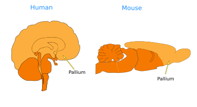
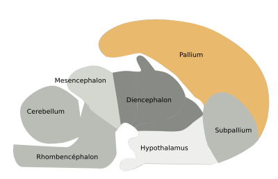
The pallium is the telencephalic region dorsal to the subpallium. In mammals, the cerebral cortical areas make up most of the pallium (Figures 1 and 2). There are four pallial areas: medial, dorsal, lateral, and ventral. In mammals, the medial pallium is the hippocampal formation, the dorsal pallium is the cerebral cortex, and the lateral pallium is the olfactory cortex. The ventral pallium encompasses part of the olfactory cortex, as well as the olfactory bulbs, claustrum, deep pallial nuclei, and the pallial part of the amygdala.


Many functions are performed by the pallium, including those superior functions that make us humans, such as learning, different types of memory, intelligence, emotions, language, social abilities, decision-making, and many others. However, it also processes primary sensory inputs, such as olfactory information, and is responsible for conscious and voluntary movements and motor planning.
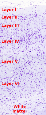
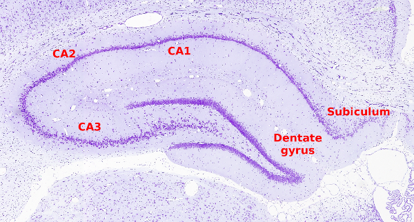
The neurons of the cortical areas are organized in layers parallel to the encephalon surface. For instance, most of the cerebral cortex (known as the isocortex or dorsal pallium) is made up of 6 layers (Figure 3). The layers I, II, and III show low neuronal density, although the neuronal content of each layer may change between cortical regions. Sometimes it is difficult to discern the border between neighboring layers, and some layers may be subdivided into sublayers. The hippocampus, a component of the medial pallium (Figure 4), and the olfactory cortex, which is the lateral pallium, contain less than 6 layers (normally 3 to 5). Cortical areas are heavily connected with areas of the same cerebral hemisphere (ipsilateral connections) as well as with the areas of the other hemisphere (contralateral connections). Contralateral connections are formed by a huge number of axons organized in axon tracts known as commissures. The most prominent commissure of the pallium is the corpus callosum, through which the dorsal cortex of the two hemispheres communicates with each other.


Besides layers, the cortical areas are organized in tightly packaged columns of neurons. The columns are oriented perpendicularly to the cortical surface. Thus, each column spans the six cortical layers. This idea of columnar organization was suggested by Lorente de Nó in 1933, and they are now called anatomical microcollums. This organization is found in all cortical regions and in most studied species.
There are two main types of neurons in the cortical areas: pyramidal and interneurons. The two types can be found in all cortical regions, although with different densities. Pyramidal neurons send axons to other cortical regions or outside the cortical areas. Interneurons project locally, that is, within the cortical layers. They are actually a rather diverse population of neurons regarding their morphology, electrophysiology, and neurochemistry.
In humans, the dorsal pallium, commonly known as the cerebral cortex, is the largest region of the pallium (and of the rest of the encephalon). It grows so much that it has to fold to get accommodated into the skull, forming the so-called convolutions, which lead the irregular surface to the human encephalon. Therefore, the cerebral cortex is divided into many regions, both anatomically and functionally (Figure 5). Anatomically, it is divided into the frontal, temporal, parietal, and occipital lobes. Physiologically, however, it can be divided into many regions, which in turn subdivide into more specialized areas. The three main cortical regions are the motor cortex (voluntary movements), the sensory cortex (sensory inputs coming mainly through the thalamus), and the associative cortex (making connections with many other cortical regions).

-
Bibliography ↷
-
Puelles L, Martínez S, Martínez de la Torre M. 2008. Neuroanatomía. Editorial Medica Panamericana S.A. ISBN: 978-84-7903-453-5.
-
 Subpalium