


The esophagus is a relatively straight tube that connects the pharynx to the stomach. In humans, it is about 25 cm long. Its histological organization shows the typical layers of the digestive tract: mucosa, submucosa, muscularis, and adventitia.
1. Anatomy
The esophagus begins at the end of the pharynx, a muscular structure to which the trachea is also connected. According to the body cavities it passes through, the esophagus is divided into three regions: the cervical, thoracic (mediastinal), and abdominal esophagus (Figure 1). The cervical esophagus begins at the pharynx and extends just behind the larynx and trachea, slightly lateral to the midline, in front of the spinal column. It measures about 4 to 5 cm. The initial portion is called the superior pharyngogastric sphincter and restricts the passage of swallowed food from the pharynx into the esophagus. The thoracic esophagus follows the cervical esophagus, passes behind the bifurcation of the trachea, and continues behind the left atrium, entering the abdominal cavity through the esophageal hiatus, an opening in the diaphragm. The thoracic exophagous measures about 20 cm. The thoracic portion can be subdivided into three further regions. The abdominal esophagus measures between 2 and 4 cm and is actually the lower esophageal sphincter, connecting the esophagus to the stomach at the cardia. Here, communication between the esophagus and the stomach is regulated, and gastric reflux is prevented.
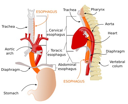
There are three constrictions that narrow the lumen of the esophagus: the upper esophageal sphincter; the point where the main bronchus and the aortic arch meet, which compress the esophagus; and the area of the lower esophageal sphincter where the esophagus passes through the abdominal wall. The greatest constriction occurs at the upper sphincter. Interestingly, there are no anatomical features that distinguish the sphincters from the rest of the esophagus, but they can be identified through pressure measurements, as the pressure is higher there.
2. Histology
The esophageal wall consists of several layers of tissue: mucosa, submucosa, muscularis, and serosa (Figures 2 and 3). The mucosa is composed of a stratified squamous epithelium, normally without a stratum corneum; however, in some animals with a diet of hard food, such as rodents, it may be partially keratinized. This epithelium transitions to simple cuboidal epithelium at the junction with the stomach. Beneath the epithelium lies the lamina media, which is a connective tissue layer containing very few cells, with scattered lymphocytes and some lymphatic nodules typically located near the excretory ducts of the mucus-secreting glands. The muscularis mucosae is, compared to other areas of the digestive tract, very thick in the upper portion, and most of its fibers are oriented longitudinally. It becomes thinner as we move toward the lower regions.
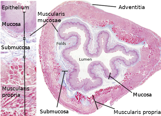

The submucosa is dense connective tissue containing a large number of elastic and collagen fibers, which allow it to stretch as the food bolus passes through. It contains numerous nerve fibers (sympathetic and parasympathetic) and sensory ganglion cells, which together form the submucosal plexus, or Meissner’s plexus. The function of this plexus is to regulate glandular secretion and the contraction of the mucosal muscle cells. The submucosa also contains a dense network of lymphatic vessels, and the mucosal lymph nodules extend into this region. The lymphatic vessels of the cervical region and the upper half of the thoracic region drain into the cervical, paratracheal, and subcarinal lymph nodes, while the lower thoracic and abdominal regions drain into the diaphragmatic, left gastric paracardial, and celiac lymph nodes. There are also numerous blood vessels, but no large vessels such as arteries are present. The secretory portions of the mucous glands that lubricate the esophageal lumen are found in the submucosa. They are more abundant in the anterior portions than in the inferior ones. The lumen of the esophagus is limited by numerous longitudinal folds of the mucosa and submucosa (Figures 2 and 4). These folds distend to allow the passage of large boluses of swallowed food.

The muscular layer consists of an outer layer of longitudinally oriented muscle cells and an inner layer of circular fibers. In the upper third of the esophagus, both layers are composed of striated (skeletal or voluntary) muscle cells. Smooth muscle fibers predominate in the lower two-thirds. In the upper region, the longitudinal layer can be divided into three fascicles that subsequently intermingle. Two of these are continuations of the pharyngeal muscles, while the third is attached to the cricoid cartilage. The cells of the circular layer form rings perpendicular to the esophageal axis in the upper and lower regions, but slightly oblique in the rest of the esophagus. This layer is slightly thicker than the longitudinal layer.
Between the two muscle layers lies a nerve plexus known as the myenteric plexus or Auerbach's plexus, composed of neurons, nerve fibers, and glial cells. This plexus innervates the muscle layers and stimulates peristaltic activity, which propels the food bolus toward the stomach.
Externally, the esophagus is covered by an adventitial layer composed of loose or fibrous connective tissue that attaches the esophagus to the rest of the body. This layer contains collagen and elastic fibers that provide structural strength. The adventitial layer is replaced by a serous layer where the esophagus joins the stomach.

Sophagus
Virutal microscopy
The esophagus contains glands that secrete mucus, whose function is to lubricate food in the upper part and protect the esophagus from stomach acid reflux in the lower part. They are divided into two types: cardiac glands and esophageal glands proper. The cardiac glands, so named because of their resemblance to those in the stomach, have their secretory portion located in the lamina propria of the mucosa and are more abundant in the lower third of the esophagus, though they are sometimes found in the upper third as well. The esophageal glands proper have their secretory portion located in the submucosa and are distributed along the entire length of the esophagus but are more concentrated in the upper half. They are tubulo-acinar glands. (see types of glands).
3. Innervation
In addition to the intrinsic innervation provided by Meissner’s and Auerbach’s plexuses, the esophagus is innervated by sensory and motor fibers of sympathetic and parasympathetic origin. The esophagus contains thermo-, chemo-, osmo-, and mechanoreceptors that collect sensory information and transmit it via the dendritic processes of neurons whose cell bodies are located in the dorsal spinal ganglia at the level of the thorax. These ganglia send their axons to neurons located in the lateral and intermediate zones of the gray matter of the spinal cord. The axons of these spinal (preganglionic) neurons can exit the spinal cord via the ventral root and make synaptic contact with ganglionic neurons of the sympathetic chain of paravertebral ganglia. The sympathetic ganglionic neurons send axons to the esophagus. There are also axons from these preganglionic cells of the spinal cord located at the T5 and T9 levels that form a nerve that travels caudally, pierces the diaphragm, and innervates the celiac ganglion. The ganglion cells of this ganglion also innervate the esophagus. All of these sympathetic fibers innervate the glands, muscles, and the esophageal enteric plexus itself, controlling its peristaltic movements.
Some of the sensory information transmitted from the esophagus is sent to brain structures, where it is interpreted as sensations (temperature, mechanical, chemical, etc.). In turn, brain structures send axons to the preganglionic neurons in the spinal cord, which modulate the activity of the esophagus.
The parasympathetic component of the esophagus is represented by innervation from the vagus nerve. Parasympathetic sensory afferents travel via fibers of the vagus nerve, which are dendrites of ganglion cells located in the inferior nodose ganglion, the jugular ganglion, and the petrosal ganglion. It has been demonstrated that some ganglia specialize in capturing information from the mucosa (chemical information) and others from the muscle (mechanical information). The axons of these ganglia terminate in various nuclei of the brainstem. The parasympathetic pathways that innervate the smooth and striated muscles of the esophagus also have different origins. Those innervating striated muscle originate from the rostral part of the nucleus ambiguus, while those innervating smooth muscle originate from the medial part of the dorsal nucleus of the brainstem. The parasympathetic pathway allows for the modulation of peristaltic activity, glandular secretion, and control of pressure in the upper sphincter. As in the case of the sympathetic system, the components of the parasympathetic system are controlled or modulated by more rostral brain structures.
4. Function
The main function of the esophagus is to serve as a passageway from the pharynx to the stomach. It is the only organ in the digestive tract where food is not absorbed. Involuntary movements of the pharyngeal muscles push food toward the opening of the esophagus, where the esophageal sphincter relaxes. Once in the esophagus, contraction waves in the esophageal wall push food toward the stomach.
5. Development
The digestive tract develops from the endoderm and mesoderm during the embryonic period. The mesoderm will form the connective and muscular tissue of the esophageal wall. By the 4th week of embryonic development, the foregut appears, from which the pharynx, esophagus, stomach, trachea, and lungs, the common bile duct, the biliary system, and the liver are formed. By the 10th week, the epithelium of the esophagus is ciliated epithelium, which is replaced by stratified squamous epithelium. However, islands of ciliated epithelium remain, which will give rise to the submucosal esophageal glands.
During the sixth week, the muscular layer and the myenteric plexus form, and by the seventh week, blood vessels invade the submucosa. The smooth muscle of the lower two-thirds derives from the somites, while the striated muscle of the upper portion derives from the pharyngeal mesenchyme.
6. Pathology
One of the most common conditions caused by impaired esophageal function is achalasia, which results from a dysfunction of the myenteric plexus cells, preventing the esophagus from moving food through due to a lack of muscle contractions. In addition, the lower esophageal sphincter cannot relax, making it difficult for food to reach the stomach.
In tumors associated with the esophagus, the absence of a serous layer allows cancer cells to spread to other tissues more easily than in other parts of the digestive tract.