

Nervous tissue develops from the embryonic ectoderm, the layer covering the embryo that becomes the epidermis. The nervous tissue is composed of two cell types: neurons and glia. The main function of nervous tissue is the processing of information coming from the external and internal environments, and then triggers a response. It is also responsible for controlling many vital functions such as breathing, digestion, heart blood pumping, regulation of blood flow, control of endocrine system, and many others. These functions mostly relay on the electrical properties of nerve cells. The stimuli are translated to the neuron language: electrical potentials that propagate through their plasma membranes. In the same way, the nervous tissue communicates with the different parts of the body, specially with the muscle cells, by electrical signals and molecules called neurotransmitters.
The majority of the nervous system is made up of cell bodies of neurons and glia, and by their cell processes. A neuropil is a nervous area with a high amount of cell processes. A scarce extracellular matrix is also present, which is enriched in glycoproteins. Extracellular matrix is involved in many functions such as cell movement, axonal growth, path finding, and formation and function of synapses.

Nervous tissue constitutes the central nervous system, including the brain and spinal cord, as well as the peripheral nervous system, including nerve ganglia, nerves and neurons scattered throughout the body.

There are parts of the central nervous system with many and tightly packed cell bodies of both neurons and glia. In fresh tissue, these areas show a grayish color, so they are known as gray matter. Other parts of the central nervous system are poor in cell bodies and are mostly made up of cell processes (mainly myelinic axons). In fresh nervous tissue, these areas are whitish, so they are known as white matter. White matter is where the larger axonal tracts are located. In the encephalon (brain), the gray matter is usually found superficially whereas in the spinal cord is found in deeper locations. Neurons are commonly associated in functional layers, as in the cerebral cortex (Figures 1 and 2) or in groups called nuclei.


In the peripheral nervous system, neurons are isolated or grouped in ganglia. The axonal and dendrite processes of these peripheral neurons, and the axons of the central nervous neurons that leaves the encephalon or spinal cord, form bundles known as nerves. They run through the body tissues and organs to send or receive information from different parts of the body.

Both the encephalon and the spinal cord are irrigated by blood vessels. The blood supply to different nervous areas can be modulated by changing the diameter of the arteries and capillaries to allow changes in the neuronal activity. Capillary diameter are regulated by pericytes. The blood flux has to be tightly adjusted because the nervous tissue is quickly damaged under low oxygen levels. Neurons die after a few minutes without oxygen. Damages caused by lack of oxygen are known as ischemia.
The nervous tissue is isolated from the surrounding tissues. Capillaries are composed of endothelial cells that form a barrier sealed by tight junctions. Furthermore, the endocytosis rate of the endothelial cells in the nervous system is lower than in other tissues. There is a layer of connective tissue, known as basal lamina, covering the endothelial cells, and wrapping the basal lamina there is a sheet of astrocytes processes, referred as glia limitans. All together, endothelium, basal lamina, and glia limitans, make the hematoencephalic barrier. This barrier controls the exchange of substances between the nervous tissue and the blood. The encephalon and the spinal cord are also separated from the bone, skull and vertebrae respectively, by membranes of tissue known as meninges.
Neurons are specialized cells in transmitting information by means of changes in the electrochemical potential of the plasma membrane. Morphologically, neurons can be divided in three compartments: cell body or soma (where the nucleus of the cell is located), axon and dendrites. The dendritic tree is the main recipient of information coming from many other neurons and sensory receptors. It integrates all the incoming information and directs the outcome to the cell body, where more integration is produced. The axon, which arises from the cell body or a primary dendrite, carries the result of the processed information to other neurons or to muscle fibers. Neurons are differentiated from the embryo ectoderm (neuroepithelial cells and neural crests) (figure 3) by a process referred as neurulation.

The number, size and organization of dendrites is variable in different types of neurons. There is only one axon per neuron (with some exceptions). Neurons communicate with each other, or with muscle cells, by using chemical mediators known as neurotransmitters. This happens in particular neuronal structures known as synapses, which consist of a presynaptic element, a synaptic cleft, and a postsynaptic element. Neurotransmitters are released by the presynaptic element into the synaptic cleft, they diffuse toward the surface of the postsynaptic element and bind to specific receptors. The binding of the neurotransmitters to receptors triggers a change in the membrane potential of the postsynaptic element, which is another neuron or a muscle cell. Sometimes, interneuronal communication is by gap junctions, known as electrical synapses. There are also neurotransmitters released outside synapses with neuromodulator roles, and the so-called neuroendocrine cells release hormones that affect other parts of the organism.

Glial cells, unlike neurons, can divide by mitosis and are as numerous as neurons in the central nervous system (Figure 4 and 5). There are several types of glial cells: astrocytes, Schwann cells, oligodendrocytes and microglia. Glia performs a large number of functions. Astrocytes wrap blood vessel of the central nervous system, cover the surface of the encephalon and spinal cord, and are present as a third element of synapses, the other two being the presynaptic and postsynaptic neurons. Although astrocytes have been regarded only as mechanical and metabolic support for neurons, they are also involved in modulating the synaptic activity. In addition, they proliferate in brain wounds and strokes, occupying the place of dead neurons. Oligodendrocytes and Schwann cells form myelin sheaths around the axons in the central and in the peripheral nervous system, respectively (see this image of myelin). Microglia is involved in functions like defense against pathogens and in damages of the nervous tissue because they act as macrophages. Microglia cells do not differentiate from the cell lineage that gives rise to neurons and other glial cells, instead they are generated in the bone marrow, and come out from blood vessels to populate the nervous tissue.

A) Dark blue labeled neurons in the striatum. The cell bodies, where the nucleus and much of the cytoplasm are located, bear thin processes that are the dendrites. In this figure, only a few neurons are stained, but there are a huge amount of neurons in this region which are not labeled. The labeled neurons contain the nitric oxide synthetase enzyme, and the activity of this enzyme produces the bluish staining.
B) Astrocytes located in the septal area showing a brown color. This color is the result of immunodetection of the glial fibrilar acid protein, a cytoskeleton protein, found in astrocytes, but not in neurons. The astrocyte morphology is visible because this protein is found in the cytoplasm.

 Muscle
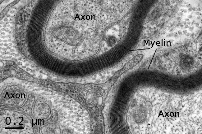