Fine features of tissues and cells are visualized with microscopes. However, only very thin samples can be readily observed with microscopes, otherwise there would be diffusion and poor penetration of light for light microscopy, or electrons for transmission electron microscopy. Because of this, it is necessary to obtain sections from tissues we want to study. Section thickness may rank from hundreds of micrometers (µm) to a few nanometers (nm). Some tissues, such as plant tissues, may be studied in thick sections (hundreds of µm). Blood cells or cell cultures are observed without sectioning because they can be extended onto a slide and form one cell-thick layer.
As mentioned in previous pages, the thinner we want the section, the harder must be the tissue before sectioning. How hard is a tissue depends on the tissue features (for example, plant cell walls make plant tissues hard) and the fixation. However, the more important step for getting hard tissues is the embedding medium. Another way for hardening samples is freezing.
Microtomes are the laboratory apparatuses for obtaining histological sections. There is a wide range of microtomes designed for different purposes: obtaining sections with different thickness, cutting soft or hard embedding media, for non-embedded tissues, and for obtaining sections from frozen samples.
Most common microtomes are as follows:
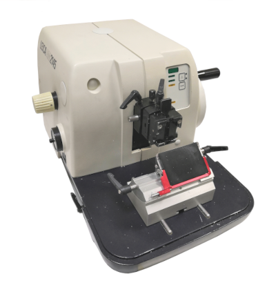
Paraffin microtome (Figure 1). It is designed to obtain sections from paraffin embedded samples. The section thickness may range from 5 to 20 µm, and they are intended for light microscopy studies. This type of microtome is present in every histology lab.

Hand microtome (Figure 2). It is a very simple and easy-to-use apparatus consisting of a sample holder that can be raised manually. The microtome is held with a hand and sections are obtained with a blade handled with the other hand. It is useful for plant samples, which have hard tissues and can be cut without embedding. Sections thickness are usually above 100 µm, intended to be observed at light microscopy.

Vibratome (Figure 3). It can cut fixed not-embedded material (relatively soft samples) and produces sections ranging from 30-40 µm to hundreds µm in thickness. These sections are studied at light microscopy.

Freezing microtome. With this microtome, 30 µm to 100 or 200 µm thick sections can be obtained from frozen samples. The sections are studied at light microscopy.
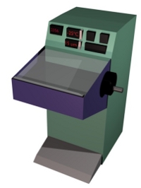
Cryostat (Figure 4). From frozen samples, 10 to 40 µm thick sections can be obtained and be studied at light microscopy.

Ultramicrotome. This apparatus is able to cut samples embedded in resin and gets sections ranging from dozens of nm to 1 µm in thickness. Thicker sections (0.5 µm or higher) are studied at light microscopy, and those thinner than 100 nm are intended for transmission electron microscopy.
Ultracryotome. It is not a widely used apparatus in histology labs, but it is used when ultrathin sections (nm in thickness) have to be obtained from non-embedded material. These samples are first frozen, therefore hardened, and then ultrathin frozen sections can be obtained for transmission electron microscopy studies.
Traditionally, the most used microtomes for the study of the general features of tissues are the paraffin microtome for light microscopy and the ultramicrotome for transmission electron microscopy. Nowadays, the cryostat is gaining popularity because it saves time (all the embedding process) and provides a general better molecular preservation. It can even cut non-fixated material.
 Embedding