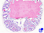
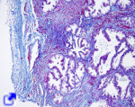
The male reproductive system consists of the testis, ducts, associated glands, and the sexual organ, or penis (Figure 1). The main function of the male reproductive system is to produce and release sperm cells for sexual reproduction. Furthermore, it is an endocrine gland that releases androgen hormones, such as testosterone, which are responsible for secondary sexual features that result in sexual dimorphism.

1. El testis
Testicles (or testis) are oval structures suspended within a bag of tissue known as the scrotum, outside the abdominal cavity. Because it is an expansion of the peritoneum, the scrotum contains two layers of mesothelium and liquid serum between them, secreted by the mesothelial cells. This aqueous solution is a lubricant that allows the movement of the testicles.
The tunica albuginea is a layer of connective tissue coating the testis, located under the tunica vaginalis. The tunica albuginea is made up of an outer layer with abundant fibroelastic connective tissue and scattered smooth muscle cells, and an inner layer enriched in blood vessels known as the tunica vasculosa. In the back part of the testis, the tunica albuginea gets thicker and forms the mediastinum testis. Several connective tissue walls, or septa, extend from the mediastinum testis to the anterior part of the testicle, dividing each testicle into pyramidal compartments known as lobules (Figure 2). Lobules are connected to each other through discontinuities in the septa. Each lobule contains from 1 to 4 seminiferous tubules, wrapped in connective tissue. There are two compartments in the lobes: seminiferous tubules and interstitial. The interstitial compartment contains smooth muscle cells, connective tissue with blood and lymphatic vessels, nerves, and interstitial cells such as Leydig cells, which produce the testosterone hormone. Macrophages are also present.


In humans, seminiferous tubules are convoluted, about 0.2 µm in diameter, and from 30 to 70 cm long. One of the ends of the tubule may be closed or connected with the tubule of another testicular lobule. They are more straight toward the posterior part of the lobule, where the seminiferous tubules from the different lobules fuse to form a testicular network known as the rete testis. In rats testis, there are about 28 seminiferous tubules in each testicle, and about 12 in mice.
The germinal epithelium forms the majority of the wall of the seminiferous tubules. It contains the male germinal cells, or spermatogonia, as well as scattered somatic cells known as the Sertoli cells. These somatic cells do not divide after sexual maturation. Germinal cells constitute most of the germinal epithelium and proliferate before and during adult life. They can divide by mitosis to produce new germinal cells or by meiosis to produce male gametes or sperm cells. Meiosis is essential for sexual reproduction because it delivers haploid cells that, after spermiogenesis, are differentiated into sperm cells.
Each seminiferous tubule is covered by a peritubular layer of fibrous connective tissue and myoid cells (smooth muscle cells). Sperm cells are produced in the seminiferous tubules and are pushed by contractions of these smooth muscle cells.
2. Ducts
The male reproductive system consists of several ducts that produce, store, and drive the sperm cells from the seminiferous tube to outside the body.
Straight segments of the seminiferous tubules form the vertices of the posterior part of the testicular lobules. The sperm cells produced in the other parts of the seminiferous tubules are collected in these segments, which are short, have no germinal cells, and whose walls are made up of Sertoli cells. The straight segments fuse to one another and form an anastomosed network of tubes known as the rete testis, surrounded by connective tissue (the mediastinum testis). The tubes of this labyrinth show a simple cuboidal epithelium.
From 15 to 20 efferent ductules leave the dorsal part of the rete testis and enter the so-called head of the epididymis, where they become very convoluted and surrounded by vascular connective tissue, forming what is known as vascular cones. In the head of the epididymus, efferent ductules converge to form the epididymis duct, which constitutes most of the next region known as the body of the epididymis. The epididymis is a very long and convoluted duct where sperm is stored. It shows a pseudostratified epithelium coated by a basal lamina and connective tissue. There is a thin layer of smooth muscle cells under the connective tissue that compresses the duct through peristaltic contractions. A layer of smooth muscle cells is also surrounded by the efferent ductules.
The epididymis becomes the ductus deferens, which drives the sperm from the scrotum to the inguinal region, coursing the lateral wall of the pelvis toward the urethra. The ductus derferens has thick walls and a narrow lumen. It is made up of a pseudostratified epithelium, a basal lamina, a thin lamina propria, and a poorly delimited submucosa. There is also a well-developed smooth muscle layer divided into three sublayers. An adventitia layer coats the muscle layer. Near the end of the ductus deferens, there is an enlargement known as the ampulla.
The ejaculatory duct is a short terminal duct that extends from the ampulla of the ductus deferens, crosses the prostate, and ends in the urethra. The epithelium of the ejaculatory duct is columnar or pseudostratified.
3. Glands
Glands associated with the male reproductive system are the seminal, prostate, and bulbourethral glands (Cowper's glands).
Seminal vesicles are long structures situated behind the prostate. Their excretory ducts join the ductus deferens and together form the ejaculatory duct. The pseudostratified epithelium of the seminal vesicles is a secretory epithelium that produces and releases the seminal fluid. This solution contains many substances, like fructose for sperm cell feeding and prostaglandins that influence the female reproductive system. The activity of the epithelium is modulated by testosterone.

Under the epithelium of the seminal vesicle, there is a layer of connective tissue highly irrigated by blood vessels. Epithelium and connective tissue constitute the mucosa. Below the mucosa, there is a profusely folded layer known as the submucosa. The crests of these folds, which are projected into the mucosa as well, may be fused at their apical parts, leaving chambers of variable sizes. Thus, the lumen of the seminal vesicle is very irregular and looks like a honeycomb when observed at low magnification. covering the inner submucosa surface, there is a layer of smooth muscle. The adventitia is the most external layer of the seminal vesicles.
The prostate is a complex gland that, in humans, consists of 30 to 50 compound tubuloalveolar glands releasing their content into the prostatic urethra. This gland is surrounded by fibromuscular stroma. The prostate is divided into four regions of different sizes: the transition zone that coats the urethra, the central zone that surrounds the ejaculatory ducts, the peripheral zone, which is the largest part of the gland, and the non-glandular fibromuscular stroma occupying the anterior part.

The prostate shows three levels of organization. There are small glands in the mucosa and submucosa of the urethra. The major glands are located in the surroundings, included in fibrous connective tissue that also contains smooth muscle cells, and altogether form the largest part of the prostate. Externally, there is a capsule of fibroelastic tissue with some smooth muscle cells. The prostatic liquid solution contains enzymes like fibrinolisins that help to decrease the viscosity of the semen.
Bulbourethral glands, or Cowper's glands, are small glands found behind the urethra. They are compound tubulo-alveolar glands that release their content into the urethra. The secretory parts consist of a cuboidal or columnar simple epithelium, surrounded by connective tissue containing striated muscle cells. The connective tissue sends tissue expansions that form septa between the secretory parts of the glands. Secreted substances are mostly lubricants, and they are released independently of ejaculation.
4. Reproductive organ
The penis is the male reproductive organ. It is organized into two dorsal columns known as corpora cavernosa and one ventral column, or the corpus spongiosum, which contains the urethra. There are connective and fibroelastic tissues covering these three structures, forming the tunica albuginea, which provides strength and support. Corpora cavernosa is a network of large anastomosed blood cavities that are filled with blood during penis erection. The cavities are coated by smooth muscle. The glans is the distal end of the penis, which is covered by the foreskin, known as the prepuce.
 Reproductive: female