1. Loose connective
2. Dense connective
2.1. Irregular
2.2. Regular
2.3. Elastic
3. Mucous connective
4. Mesenchima
5. Reticular
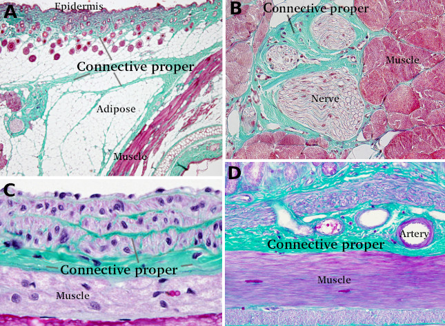
The Connective tissue proper contains several types of cells and a variable amount of extracellular matrix made up of fibers and ground substance (Figure 1). This tissue is widespread throughout the body. It fills spaces between organs (Figure 2), for example between the skin and muscles, and surrounds blood vessels, nerves and several organs. It constitutes also the part of the stroma of organs such as the kidney, liver, glands, gonads, and some others. Connective proper forms tendons, ligaments, cornea and dermis.
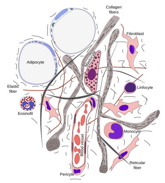
The typical cell type of connective tissue proper is the fibroblast (Figure 3). Its main function is to synthesize and released the majority of molecules that form the extracellular matrix. At light microscopy, fibroblasts are elongated cells, more or less fusiform or irregular in shape, with one ovoid nucleus containing one or two nucleoli, and generally showing small amount of cytoplasm. Other cells, such as reticular and mesenchymal cells, can be found in particular subtypes of connective tissue proper (see below).
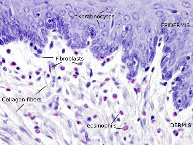
Fibroblasts are regarded as resident cells because they are typically found in the connective tissue proper. Other cells are originated in the bone marrow and reach the connective tissue proper after crossing the endothelium of blood vessels. These non-resident cells are mast cells, macrophages, plasmatic cells and the different types of leukocytes, which are intermingled with fibroblasts. All these non-resident cells are involved in immune functions and are able to move through the extracellular matrix. The amount of non-resident cells is variable depending on the location and the local conditions. Adipocytes are also present in connective tissue proper. It is interesting that adipocytes and fibroblasts share the same mesenchymal precursor.
Considering the amount and molecular composition of the extracellular matrix, and the cell types, there are several subtypes of connective tissue proper.
1. Loose connective tissue
Elastic fibers.
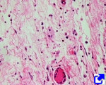
Areolar or loose connective tissue is the most abundant connective tissue proper, characterized by an irregular organization of abundant and not dense extracellular matrix containing scattered cells. It shows a broad distribution since it can be found almost in every organ of the body, both in the inner and outer parts. It occupies regions which are not under strong mechanical forces. Loose connective tissue fills spaces between the skin and muscles, is found under many epithelial tissues, wraps many organs, it is part of the stroma of organs like the kidney, livers, testis, and many others, and forms the walls of the digestive tract.
Loose connective tissue is mainly composed of fibroblasts and abundant extracellular matrix. The extracellular matrix contains scattered collagen and elastic fibers, and much less abundant reticular fibers. This tissue plays a fundamental role in nurturing other tissues and organs since nutrients easily diffuse through the ground component of the extracellular matrix. There is a dense network of blood vessels, nerve processes, as well as secretory parts of exocrine glands. It is not a specialized tissue.
2. Dense connective tissue
In the extracellular matrix of dense connective tissue, the collagen and elastic fibers are more abundant than the ground component. Thus, unlike loose connective tissue, there are not so many clear spaces. The fibroblasts of dense connective tissue are called fibrocytes to remark that the activity is much lower than in the loose connective tissue. There are also lower cell type diversity. Some times it is not so easy to distinguish between loose and dense connective tissues because there are regions with intermediate features between both tissue types. The main function of dense connective tissue is to withstand mechanical forces. There are three subtypes of dense connective tissue: irregular, regular and elastic.
2.1. Irregular
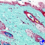
Dense irregular connective tissue has large amounts of collagen fibers grouped in thick bundles forming a tridimensional network. It is a strong mechanical tissue. Collagen fibers are thicker and more numerous than in the loose connective tissue, and there are less complex networks of blood vessels and nerve fibers. This tissue forms the dermis (mostly the reticular dermis) and in capsules around organs, dura mater meninges, periosteum, pericardium, cardiac valves and articular capsules.
2.2. Regular

Dense regular connective tissue contains a large amount of collagen fibers in the extracellular matrix arranged in parallel bundles or sheets. This reflects mechanical needs. Actually this tissue is found in structures under unidirectional mechanical stress, such as tendons, ligaments, and sheaths or fascias surrounding skeletal muscles. Dense regular tissue is also found in the fascia (aponeurosis) of the abdominal muscles, where fibers are oriented in different directions because stretching are coming from different directions. The cornea is another structure made up of dense regular connective tissue with layers of collagen fibers oriented perpendicularly between each other.
2.3. Elastic
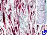
Elastic connective tissue is characterized by the abundant elastic fibers, which provides a high elasticity to the tissue, as well as a yellowish color. This type of tissue is found in those organs under high mechanical stress(stretching and contractions) caused by pressure or stretching. Elastic fibers are usually arranged in bundles of variable thickness, but can also be found as single fibers. Loose connective tissue is often found surrounding the elastic connective tissue providing cohesion. Elastic connective tissue is found in elastic ligaments that join vertebrae allowing bending of the vertebral column. Other locations are the ligament of the nape and the short ligaments of the larynx.
3. Mucous connective
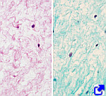
Mucous (also known as gelatinous) connective tissue looks like gelatin. It is highly hydrated, turgid, and show high mechanical resistance. The proportion of extracellular matrix may be up to 95 %. Mucous connective contains a low number of cells with myofibroblast-like features. The most abundant proteins of the extracellular matrix is type I collagen, which forms thin fibers. Proteoglycans such as chondroitin sulfate and dermatan sulfate are abundant, as well as hyaluronic acid.
This tissue is found during the embryonary period, but it is rare in adults. It is the main component of the umbilical cord, where it forms a spiral structure called Wharton jelly. Unlike other tissues, only the myofibroblast cell type has been found in the human mucous connective tissue. It lacks blood and lymphatic vessels, excepting two arteries and one vein that communicate the embryo with the placenta. This tissue is also found in the chorionic plate of the placenta, around fetal capillaries, and in the crest of chickens.
The mucous tissue of the umbilical cord is under heavy research because their cells may become pluripotent stem cellsthat can be differentiated in many cell types. It makes this tissue a potential source of cells for regenerative therapies and tissue engineering.
4. Mesenchyma
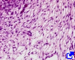
Mesenchymal connective tissue is composed of undifferentiated cells and loose extracellular matrix. The extracellular matrix allows a large motility of mesenchymal cells, very useful to organize and form new structures during the embrionary period. It can be regarded as a transient tissue because it is abundant in the embryo, but scarce in the adult. However, it is found in the bone marrow, fat depots, muscles, and milk teeth dental pulp. In these tissues, mesenchymal cells are called mesenchymal stem cells.
During embryo development, mesenchymal tissue differentiates from mesoderm. However, there is mesenchymal tissue in the head derived from the neural crests. Many other tissues are formed from mesechymal tissue: other connective tissues, cartilage, bone, the majority of blood and lymphatic systems, as well as some smooth muscle. In addition, signals sent from mesenchymal tissue initiate many body organs and structures.
5. Reticular
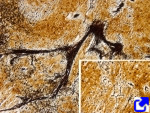
Reticular connective tissue is characterized by having abundant reticular fibers. Cells synthesizing these fibers are fibroblasts called reticular cells. Reticular fibers, or reticulin, may be present in other connective tissues, but they are more numerous in the reticular tissue. They are composed of type III collagen, that makes thinner (around 150 nm in diameter), branched and anastomosed fibers, containing a high proportion of carbohydrates. Branching is a feature of reticular fibers, unlike other collagen fibers, like those formed by types I and II collagen. They are so thin that are not visible with the light microscopy unless specif staining is performed, such as silver impregnation or PAS histochemistry.
The main role of reticular connective tissue is to provide a scaffold for mechanical support of several types of cells, as well as reticular cells. There are many cells like lymphocytes, adipocytes, smooth muscle cells, macrophages, hematopoietic cells, and some others. Thus, cell density is higher than in other connective tissues. Reticular cells are usually linked to reticular fibers forming a relatively stiff scaffold, whereas the other cells can move more freely. The function of the reticular connective tissue is important in organs like lymphoid nodules, kidneys, artery wall, spleen, liver, bone marrow, tonsils, and Peyer plates of the ileum It is also sparsely present in other body organs.
-
Bibliography ↷
-
Bibliography
Davies JE, Walker JT, Keating A. 2017. Concise review: Wharton’s jelly: the rich, but enigmatic, source of mesenchymal stromal cells. Stem cellls translational medicine. 6: 1620-1630
.
Krstić, R. V. 1989. Los tejidos del hombre y de los mamíferos. Editorial Interamericana. McGraw-Hill. Madrid. ISBN: 84-7615-413-5.
MacCord, K. Consultado en 2017. Mesenchyme. The Embryo Project Encyclopedia.
-
 Connective tissue