

Blood is regarded by many authors as a specialized type of connective tissue composed of cells, cell fragments and a liquid extracellular matrix known as blood plasma. Blood is the liquid found inside blood vessels and heart. Heart beating and body movement push the blood through the cardiovascular system, which reaches every part of the body. The volume of blood in the human body depends on the body size. A 70 kg body contains around 5 to 6 liters of blood. The blood temperature is about 38 ºC, one degree higher than the general body temperature. This higher value is a consequence of the friction of the blood inside the blood vessels, mainly in the small diameter blood vessels.
Blood has many functions. The following are three salient functions. 1) Communication pathway. Blood transports nutrients and oxygen from intestine and the lungs, respectively, to the rest of the body. Wasting products are transported to the kidney and lungs. It is also the main communication pathway for chemical signals, like hormones, between distant cells in the body. 2) Homeostasis. Blood contributes to the general body homeostasis. For example, it keeps relatively constant the body temperature and pH of tissues. 3) Defense. Blood contributes in the repairing of wounds by sealing the damages with erythrocytes, platelets and plasma, i.e., blood clotting. It also contains the cells of the immune system, that use the circulatory system to be transported and attack pathogen in any tissue of the body.
1. Blood cells
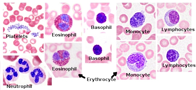
Blood cells are classified in two groups: erythrocytes or red cells, and leukocytes or white cells (Figures 1 and 2). There are also cell fragments in the blood referred as platelets. Leukocytes can be granular leukocytes: neutrophils, basophils and eosinophils, and agranular: lymphocytes and monocytes. Most blood cells are erythrocytes (99% of the cells). All blood cells develop from a common stem cell that, in adult animals, is found in the bone marrow.

The different components of the blood can be separated by density gradient centrifugation. The heaviest elements are erythrocytes, that fall to the bottom of the centrifugation tube. Leukocytes and platelets are found a little higher forming a whitish layer. Plasma is the lightest component and remains in the superficial part of the centrifugated. In human males, blood contains 47 % of eyrhtrocites, while in human females is about 41 %. The proportional volume of eythrocytes out of the total blood volume is known as hematocrit. The proportion of leukocytes is lower than 1 %. Plasma constitutes the rest of the blood. The red color of blood is the result of the high content in hemoglobin of erythrocytes, which is darker when the oxygen level is low. The blood serum is the plasma without the blood cotting agents.

Erythrocytes give the red color to the blood because of its high hemoglobin content, a protein containing iron in its structure. The main role of erythrocytes is transporting O2 and CO2. In mammals, erythrocyte can be regarded as a highly modified cell for this function because it has no nucleus and lacks mitochondria and other cellular organelles. It is shaped as a biconcave disc of about 7.5 µm in diameter, which renders a larger exchange surface in contact with the blood plasma.

Platelets, or thrombocytes, are small portions of cytoplasm without nucleus. At light microscopy, they can be observed as small structures of about 2 to 5 µm in diameter, colorless or slightly basophilic. They contain some internal membranous compartments like dense specific azurofilic granules, mitochondria (one or two per platelet), and clear vesicles and tubules. They also contain glycogen granules. The platelet main function is to cooperate during agglutination and blood clotting. They are present in mammals, but not in lower vertebrates. Platelets are generated by fragmentation of the cytoplasm of megakaryocytes, a cell type found in the bone marrow.
Leukocytes (white blood cells) are nucleated cells and colorless in fresh blood. Their main function is to defend the body against external aggression such as pathogens and against malfunctions and alterations of the body tissues. These functions are performed outside the blood stream, as they have the ability to cross the wall of blood vessels and act in the damaged tissues. Actually, they use the circulatory system to travel through the body. Leukocyte cytoplasm contains two types of granules: azurophilic or primary granules, which are lysosomes, and specific or secondary granules containing diverse types of substances. Leukocytes are classified into granular and agranular. They all have azurophilic granules, but the specific granules only are found in the granular leukocytes.
Granular leukocytes are neutrophils, eosinophils and basophils. Agranular leukocytes include lymphocytes and monocytes. Neutrophils are the most abundant granular leukocytes and account for 60-70% of the total leukocytes. They are easily recognized by their multi-lobed nucleus and show abundant specific granules and some cytoplasmic azurophilic granules. Specific granules contain lysozymes, complement activators, collagenases, and other enzymes. They are very important during the defense against bacterial infections. Eosinophils are up to 2 to 5% of the leukocyte population. Their nucleus is bi-lobed and their cytoplasmic specific granules show a strong affinity for acid dyes such as eosin. These granules have basic proteins such as major basic protein and eosinophil cationic protein, which are involved in the control of parasitic infections, and histaminases that neutralize the action of histamine in allergic reactions. Basophils are the less abundant and smaller granular leukocytes, accounting for 0.5% of the total leukocytes. Its nucleus is slightly lobed. They contain specific granules which are stained by basic dyes such as haematoxylin. The cell membrane of basophils has receptors for immunoglobulin E, and the specific cytoplasmic granules contain histamine and heparin. Thus, it is suggested that these cells act at the connective tissue cooperating with mast cells.
Agranular leukocytes lack specific granules in the cytoplasm, but they have a small population of nonspecific granules. After neutrophils, lymphocytes are the second most abundant type of leukocytes, accounting for 20 to 35% of the total leukocytes. They are small and show some size variability, apparently not related to the different types of lymphocytes. The two main groups of lymphocytes are B and T. Both are major players in the immune defense of the organism. Monocytes are the other agranular leukocyte type. Monocytes show large size in blood smears and have a kidney-shaped nucleus. They help in the defense of the body by leaving the blood and moving to the site of infection or injury, where they become macrophages.
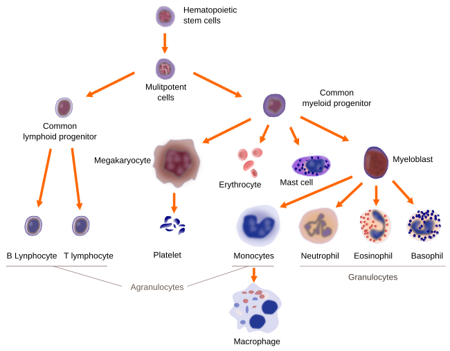
Generally, the lifespan of the cellular components of the blood is very short, ranging from a few hours to a few weeks (excepting some memory lymphocytes that can live for years). Therefore, blood cells are produced continuously by a process known as hematopoiesis (Figure 3). In humans, hematopoieses takes place in different organs during development: in the viteline sac in embryos, in the liver, spleen and lymphatic tissues in fetuses, and then in the red bone marrow. After birth, hemaopoietic process is moved to the bone marrow of long bones. In adults, the main hematopoietic centers are bones: cranial bones, pelvis, vertebrae, sternum, and regions near the epiphisis of the femur and humerus. Under some circumstances, hematopoiesis may be initiated in the adult liver and spleen.
2. Plasma
Plasma is the liquid part of the blood and accounts for more than half of the blood volume. It is 90 % water, and the rest is composed of proteins, ions, amino acids, lipids, and gases. Plasma is the main transporter of nutrients and waste products.
Albumin is the most abundant protein in the plasma (54 % of the total protein content) and perform several functions. Many molecules associate with albumin to be transported through blood stream, such as fatty acids and steroid hormones. Albumin is also the main factor maintaining the blood pressure, which in turn regulates the blood volume. Globins are the second most abundant proteins in the plasma. They are a group of proteins accounting for about 38 % of the plasma proteins, and can be divided in three types: alpha, beta and gamma. Alpha and beta globins are synthesized in the liver and carry iron, lipids, and liposoluble vitamins. They also contributed to the plasma osmolarity. Gamma globins or gamma globulins are the soluble antibodies of the immune system, also known as immunoglobulins. Fibrinogen is another protein of the plasma. It is not very abundant, but very important for blood clotting. Fibrinogen is synthesized in the liver. Proteins that form the plasma may be specific of the plasma or may be also present in other tissues, like enzymes, immunoglobulins and hormones.
 Bone
Bone 