Together with connective, muscle, and nervous tissue, epithelium is one of the four basic types of tissues in the animal body. It accounts for more than 60 % of the total cells of the human body. Epitehlium lines the internal cavities and external surfaces of the body. In addition, epithelial derivatives constitute the main secretory cells of the organism, and in some cases, like the liver, they form the organ parenchyma.
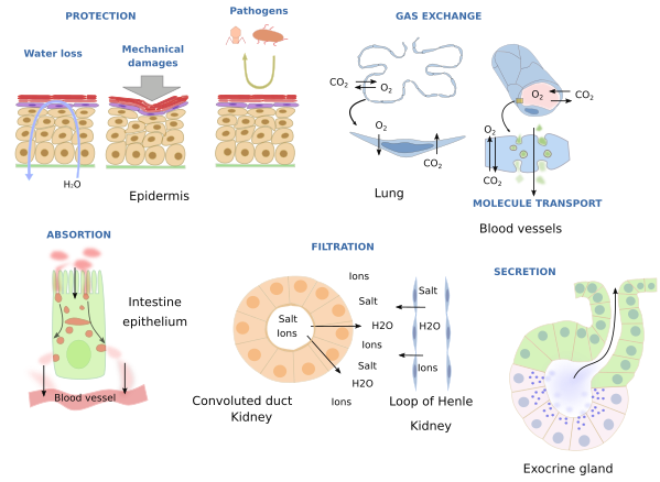
Many functions are accomplished by epithelium: protection against mechanical assaults, prevention of water loss, filtering, selective absorption, secretion, exchange of gas and other molecules, substance transport over their surfaces, and may contain cells that work as sensory cells (Figure 1).

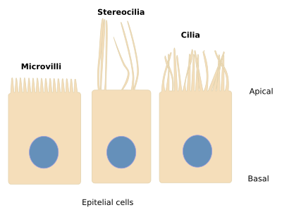
Some of the aforementioned functions are carry out with the help of the specializations of apical domain of the cell: microvilli, stereocilia and cilia (Figure 2).

Epithelial tissues are made up of tightly joined cells and show large cell-cell contact surfaces that leave very little extracellular matrix. Several molecular complexes form these cell-cell junctions, such as tight junctions (zonula occludens), desmosomes (zonula adherens), and adherent junctions (zonula adherens). Tight junctions establish strong cell-cell adhesion and get plasma membranes of neighboring cells so close between each other that the extracellular space is very narrow or nearly occluded. Desmosomes and adherent junctions are more abundant. Cadherins mediate these two last cell junctions, connecting the cytoskeleton of adjoining cells and providing cohesion and strength to the whole epithelium. Cell adhesion may be modulated, either reinforced or weakened, depending on the physiological needs. Cytokeratins are the typical intermediate filaments of epithelial cells.
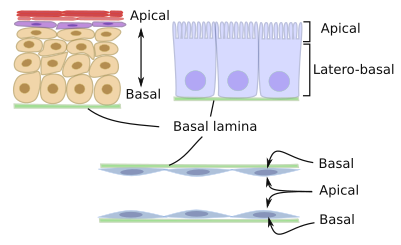
Epithelial cells are organized in one or several strata that rest on a specialized sheet of extracellular matrix called basal lamina. The basal lamina is a highly organized layer of extracellular matrix that completely covers the epithelial basal surface. It is produced by both epithelial cells and the underlying connective cells. Polarization is another feature of epithelial tissues. It means that they perform different functions in the apical domain (facing the lumen of the organ or the exterior of the body) compared with the basal domain (in contact with the basal lamina) (Figure 3). Polarity is reflected in the epithelial the cell morphology, particularly when the epithelium is a single cell layer. In this case, it is said that cells have an apical and a basal domain.

Epithelial tissues do not have a blood capillary net (except the stria vascularis of the inner ear). So, they get food by diffusion from the underlying connective tissue. Nutrients need to cross the basal lamina.
En general, epithelial tissues are composed of a more abundant cell type, and by other less numerous cell types. For example, enterocytes consitute the majority of the intestine epithelium, but caliciform, Paneth and enteroendocrine cells are also present. In the same way, the majority of the epidermis is made up of keratinocytes, but more scarce melanocytes and Langerhans cells are found too. The trachea epithelium consists of at least 6 cellular types. However, some epithelial tissues, such as endothelium, might be formed by only one type of cells, although it looks like that endothelial cells form a heterogeneous population.
Epithelial tissues show a high rate of renewing and regeneration. This is particularly high in those epithelia exposed to the external environment, like epidermis, digestive epithelium and respiratory epithelium. The proliferation of epithelial cells happens all the time, but it is increased when some wound needs to be repaired. Adult stem cells are present in epithelial tissues, and are usually found in the basal part, contacting with the basal lamina. They proliferate and differentiate into most of the cell types of the epithelial layer.
It could be thought that epithelial cells are non-moving cells because of the strength and high number of cell adhesions between each other. It is not so in some epithelial tissues. Cell adhesion complexes are dynamics, they can be formed and disassembled, which makes possible cell movements. In these cases, the epithelium behaves as a fluid. This feature allows new cell recruitment after proliferation, replaces cells that die by apoptosis or that are extruded, increases the surface of the epithelial layers during development. All these processes need to be done without losing the epithelial integrity, and therefore the barrier function keeps working. Epithelial tissues have the ability of "sensing" mechanical stimuli, so that when an epithelium is stretched the proliferation rate is increased. Mechanical forces are detected by membrane receptors that are activated when the cell is stretched, allow the entrance of calcium into the cell, and begins a signaling cascade that activates B cyclin, a molecule that favors the cell cycle. On the other hand, when stretching disappears there is an inhibition of the cell cycle. It means that epithelial cells "sense" if mechanical stimuli are in a proper range. Proliferation is inhibited if mechanical stresses are below this range, and it is favored when it is above. Epithelial cells can move laterally in the epithelial layer to counteract these mechanical forces and to get distributed properly.
Epithelium receives different names depending on where it is located. For example, the skin epithelium is referred as epidermis, the epithelium lining the internal surfaces such as abdominal cavities or cardiac cavity is known as mesothelium, and blood vessels are internally lined by the epithelium known as endothelium. Furthermore, epithelium can be classified according to the number of cell layers (simple or stratified) and the shape of the most superficial cells (squamous, cuboidal and columnar). Epithelium may have apical structures such as microvilli, cilia and flagella. The embryonary origin of epithelia may be followed to the three germ layers: endodermo, ectoderm and mesoderm: epidermis differentiates from ectoderm, endothelium from mesoderm, and digestive epithelium from endoderm. Some epithelial tissues like epidermis may differentiate and organize their cells to produce macroscopic structures such as hair, nails and feathers. These structures are induced by the underlying mesoderm.
In addition, the epithelium that cover the body surfaces in called covering epithelium. Some epithelial cells may become specialized in secretion of a wide diversity of substances, and they may be organized as glands. They constitute the glandular epithelium. The secretory units of glands are wrapped by myoepithelial cells, which have the ability of contraction and are differentiated from epithelial cells.
There are some very specialized epithelia with functions not related to covering or secretion. They are grouped under special epithelia. Neuroepithelium (olfactory and gustatory epithelium), germinal epithelium (it forms seminiferous tubules of testis) and myoepithelial cells (with contractile ability) are among these special epithelia.
 Introduction