Covering epithelia are sheets of tissue that cover the external surfaces (skin, lungs, gut) and line the internal cavities (blood and lymphatic vessels, pleura) of the body. Their major function is as barrier between exterior and the interior of the body, and between two internal environments, like blood and other tissues. Epidermis is an epithelium covering the external surface of the body. It provides protection against pathogens or mechanical damages, prevent water loose, and perform many other functions. Pleura is a type of covering epithelium known as mesothelium. It coats the serous cavities of the body and internal organs. Endothelium is a type of covering epithelium that lines the interior surfaces of blood and lymphatic vessels. Covering epithelium shows almost no extracellular matrix and epithelial cells are tightly attached to one another by macromolecular adhesion complexes. Some epithelia show a high rate of cell turnover where cell death and cell proliferation are frequent. Some epithelial cells can have apical specializations that allow them to function as sensory receptors, and some animals show complex structures in their epithelial layer, such as hair, feathers or scales.
Epithelia are usually classified according to two features: the number of cell layers and the shape of the cells of the more superficial layer (Figures 1 and 2). Simple epithelia are single cell layers where all the cells contact the underlying basal lamina and have an apical free surface. The shape of the cells can be flat (wider than high), cuboidal (as wide as high), or columnar (higher than wide). Pseudostratified epithelia are simple epithelia where all cells contact the basal lamina, but not all cells reach the superficial layer because some are shorter than the others. Thus, this is a simple epithelium that looks stratified. Stratified epithelium contains two or more layers of cells. Only cells of the deeper layer are in contact with the basal lamina and only cells of the upper layer show free surfaces. Stratified epithelia can be classified as squamous, cuboidal and columnar, depending on the shape of the cells of the upper layer when observed in transverse view. Transitional epithelium is another type of stratified epithelium that can be stretched, changing the shape of its cells.
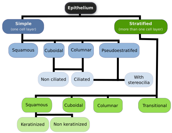
Squamous Cuboidal Columnar
Cell arrangement
Simple Stratified Pseudostratified Transitional
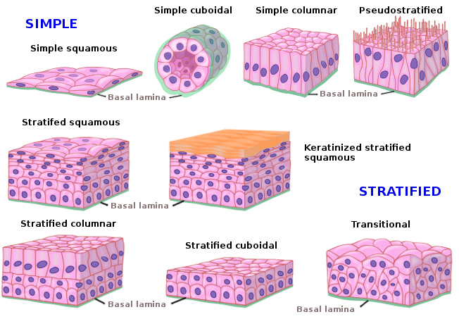
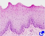
Squamous epithelium
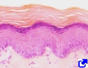
The outer layer is formed by flat cells.
Cuboidal epithelium
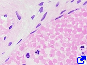
The outer layer is formed by cube-like cells.
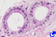
the excretory duct of a gland.
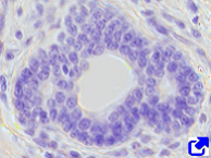
Columnar epithelium
The outer layer is formed by columnar shaped cells
Transitional epithelium
It can be streched. The cell shape changes during these movements.

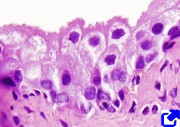
Pseudostratified epithelium
It contains cells at different levels, but all of them contact with the basal lamina. That is why this epithelium looks like stratified.


 Epithelium