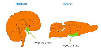
The hypothalamus is a relatively small region topographically found ventral to the prethalamus and thalamus (Figures 1 and 2). That is why the name hypothalamus and why it has been traditionally included as part of the diencephalon. However, that position is the result of the bending of the neural tube during embryonic development. Actually, topologically, the hypothalamus is rostral to the diencephalon, more precisely to the p3 diencephalic region. In fact, together with the telencephalon, the hypothalamus is the most rostral part of the neural tube. The hypothalamus and the telencephalon constitute a segment called the secondary prosencephalon (Puelles and Rubenstein, 2015, suggested the name "hypotelencephalon"). The hypothalamus is a structurally complex region with 33 subregions that may give rise to more than 150 different neuronal populations.


The hypothalamus is divided into an alar plate and a basal plate. Rostrocaudally, it is divided into a rostral, or terminal, hypothalamus (hp2) and a caudal, or penduncular, hypothalamus (hp1) (Figures 2 and 3). The border between both extends until the telencephalon. The rostral hypothalamus encompasses the supraoptic, lateral anterior, suprachiasmatic, anterior, anterobasal, ventromedial, arcuate, and mammillary nuclei. The neurohypophysis is found in its basal region. The rostral hypothalamus is dorsally adjacent to the preoptic area. The caudal hypothalamus contains the paraventricular nucleus, part of the dorsomedial nucleus, and the retromamillary area. Dorsally, it is limited by the telencephalon.

Functions
The hypothalamus is a relatively small area in the vertebrate telencephalon, but it performs essential functions for the organism, such as the control of hunger, thirst, circadian rhythm, body temperature, reproduction, sexual behavior, emotions, and so on. Unlike other nervous regions that communicate with the body through motoneurons innervating muscles, the hypothalamus releases hormones into the blood vessels. This happens mostly at the neurohypophysis, but also at the median eminence. In this way, the hypothalamus is a major regulator of the endocrine system. There are two main neuronal populations that release hormones: magnocellular and parvocellullar neurons. The magnocellular neurons of the paraventricular and suprachyasmatic nuclei release oxytocin and arginin-vasopressin through the axons that reach the neurophypophysis. The parvocellular neurons are distributed in the tuberal, preoptic, arcuate, and anterior periventricular and paraventricular nuclei. They project to the median eminence and release several types of hormones. Anyway, the release of hormones occurs after the integration of different types of information coming from other encephalic regions and sources. Visual, taste, smell, and light stimuli are included in these inputs.
Reproduction
The male and female reproductive cycles are modulated by the interaction of many hormones released from different endocrine glands, including the hypothalamus, hypophysis, and gonads. The hypothalamus releases the gonadotropin-releasing hormone (GnRH) into the anterior hypophysis (adenohypophysis), which in turn releases the luteinizing hormone (LH) and the follicle-stimulating hormone (FSH) into the blood stream. These hormones are released in both males and females and stimulate gamete production and maturation of the ovarian follicles.
Oxytocin is produced by the magnocellular and parvocellullar neurons of the paraventricular and supraoptic nuclei of the hypothalamus, and it is transported through their axons to the posterior hypophysis (neurohypophysis), where it is released into the blood. In females, oxytocin leads to uterus contractions at the time of birth and to the production of breast milk. These actions are mediated by oxytocin receptors (OTR), which are also expressed in several encephalic regions, such as the hippocampus, neocortex, amygdala, and accumbens nucleus. During pregnancy, the medial nucleus of the hypothalamus increases the expression of oxytocin. This action of oxytocin on different encephalic regions activates maternal behavior.
Diuretic control
Molecules regulating the liquid content of the body are released from the hypothalamus. Vasopressin is a hormone that controls the water content of the body. It is synthesized in the magnocellular neurons of the suprachiasmatic and paraventricular nuclei of the hypothalamus. It is also released by the parvocellular neurons of the paraventricular nucleus. The magnocellular axons release vasopressin into the blood vessels of the posterior hypophysis. Vasopressin is also known as the antidiuretic hormone because it increases the uptake of water in the kidneys and induces vasoconstriction. The receptors that sense the amount of water in our body are osmoreceptors found in the hypothalamus.
-
Bibliography ↷
-
Bibliography
Diaz M, Morales-Delgado N, Puelles L. 2015 Neuroanatomía. 2015. Ontogenesis of peptidergic neurons within the genoarchitectonic map of the mouse hypothalamus. Frontiers in neuroanatomy. 8:162.
Puelles L, Martínez S, Martínez de la Torre M. 2008. Neuroanatomía. Editorial Médica Panamericana S.A. ISBN: 978-84-7903-453-5.
Puelles L, Rubenstein JLR. 2015. A new scenario of hypothalamic organization: rationale of new hypotheses introduced in the updated prosomeric model. Frontiers in neuroanatomy. 9: 27.
-
 Diencephalon