The digestive system shows a variety of organizations in both invertebrates and vertebrates. In vertebrates, the digestive system is a duct connecting the mouth, in the rostral part of the body, with the anus, in the caudal part of the body. Although there are major differences when comparing the digestive systems between species, mostly due to the type of food they eat, we are going to describe the general organization of the digestive system of mammals with an omnivorous diet.
The digestive system can be divided into a cephalic region, an axial part, and two large glands: the liver and pancreas (Figure 1).

The cephalic area includes the oral cavity, salivary glands, and teeth, as well as the tongue, and the pharynx, which connects the digestive and respiratory systems. The oral and nasal cavities are separated by the palate, except at the pharynx level. Both the hard palate (anterior) and the soft palate (posterior) are covered by the same type of epithelium as the rest of the oral cavity: stratified squamous epithelium. When comparing different animal species, the cephalic part shows the higher structural variability of the digestive system. The functions of the cephalic area are mechanical digestion, secretion of digestive enzymes like amylase, swallowing, as well as taste sensing.
The oral cavity is connected with the esophagus through the pharynx. The pharynx is involved in both digestion and breathing. When food enters the pharynx, the ducts that drive the air to the lungs are closed. The pharynx can be divided into an upper part related to the nasal cavity, the middle oropharynx region, and the caudal nasopharynx, which is related to both digestion and breathing.
The axial part of the digestive system comprises the esophagus, stomach, and intestines (small, large, and rectum). The ingested food goes through the esophagus to the stomach. In the stomach, the low pH and degradation enzymes produce partial digestion. After that, food moves to the small intestine for further breakdown of the meal. Here, water and nutrients are absorbed through the epithelium and transferred to the blood and lymphatic vessels. Finally, leftover products are pushed into the large intestine and defecated through the anus.
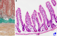
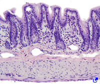
The histological organization of the esophagus, stomach, and intestine is in four layers (Figure 2). The mucosa is the most internal layer, facing the lumen. It consists of an epithelium that covers the internal surface of the digestive tube, showing different morphological features related to protection, secretion, and absorption functions performed along the digestive tube. The epithelium rests on the basal lamina, and the basal lamina rests on the lamina propria. The lamina propria is highly irrigated, smooth connective tissue containing many cells of the immune system, like macrophages, plasma cells, and lymphocytes. The epithelium, basal lamina, and lamina propria form the mucosae. In the lamina propria, there is a discontinuous layer of smooth muscle cells known as the muscularis mucosae, found in the outermost part of the mucosa (far away from the epithelium). The muscular mucosae makes possible the movement of the mucosa.
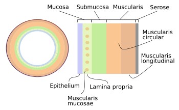
The submucosa is the irregular, dense connective tissue surrounding the mucosa. It contains many exocrine glands, known as submucosal exocrine glands. It is also highly irrigated by blood vessels and innervated by a dense network of nerves that form the Meissner's plexus, which controls the motility of the mucosa and the secretory activity of the exocrine glands.
The muscularis, or muscle layer, covers the submucosa. It is made up of smooth muscle cells, except for the upper part of the esophagus, wich shows skeletal striated muscle fibers. This muscle layer is organized into two sublayers, an inner layer with circularly arranged cells and an outer layer with longitudinally arranged cells. Between both layers, there is a neurovegetative neural plexus, known as Auerbach's plexus, that drives the muscle contraction, resulting in peristaltic movements along the digestive tube.
The serose, is the outer layer. It is made up of loose connective tissue with many adipose cells. This layer sets the border between the digestive tube and the coelomic cavities, and it is dorsally attached to the mesentery.
Two major glands release their secretory products into the digestive duct: the liver and pancreas. Both share the bile duct, the common bile duct, as an excretory duct that ends up in the small intestine. The liver releases bile fluid containing biliary acids needed for digestion and the absorption of fat. The exocrine part of the pancreas releases many enzymes for digestion. Both glands also have endocrine functions by releasing hormones into the blood. The hepatocytes of the liver are able to synthesize and release both exocrine and endocrine compounds, whereas the endocrine and exocrine functions are performed by different parts of the pancreas. The Langerhans islets are the endocrine structures of the pancreas.


 Reproductive: male