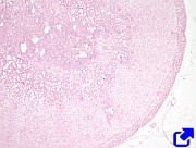
1. Hypothalamus
2. Hypophysis
3. Pineal gland
4. Thyroid
5. Parathyroid
6. Suprarrenal
7. Gonads
8. Thymus
9. Pancreas
10. Others
The common feature shared by the structures of the endocrine system is the synthesis and release of molecules known as hormones. Hormones are released into the extracellular milieu and reach the blood stream to be transported everywhere in the body. However, some hormones may perform their functions locally. Hormones carry information to the target cells, influencing their activities. The final effect depends on the hormone, the target cell type, and the physiological state of the organism. The response of the body to hormones is commonly slower than to other chemical signals, such as neurotransmitters in the nervous system or in motoneruon-muscular cell transmission.
Hormones can be defined as molecules that induce target cell to perform a function. More than 100 hormones have been identified in humans. Chemically, they can be classified as steroids, proteins, and those derived from amino acids. Each hormone is detected by specific receptors found in the target cell. These receptors can be located in the plasma membrane, where they recognize peptidergic and catecholaminergic hormones and initiate intracellular signaling pathways, resulting in, for instance, an increase in cAMP. Other hormones, such as thyroids and steroids, can easily cross the plasma membrane and are detected by intracellular receptors, which contain a molecular domain that allows them to bind to the DNA and therefore regulate gene expression.
The cells that synthesize and release hormones are known as endocrine cells. They can be found throughout the body as single cells in organs like the gonads, digestive ducts, and liver. These endocrine cells are together known as the diffuse endocrine system. Other endocrine cells associate to form glands, referred to as endocrine glands, such as the hypophysis, pineal gland, thyroid gland, parathyroid gland, and adrenal glands. The major structure that regulates the endocrine system is the hypothalamus, found in the rostral telencephalon (ventral position). The endocrine glands are heavily irrigated by blood capillaries. In these glands, the endocrine cells are arranged into cords or islands, and sometimes in follicles, as in the thyroid gland.
1. Hypothalamus

The hypothalamus is an intermediary structure between the central nervous system and the endocrine system. It releases hormones that inhibit or promote the release of hormones by other endocrine cells and glands in the body. In this way, the hypothalamus promotes body homeostasis by regulating the heartbeat, blood pressure, appetite, digestive secretions, the activity of other glands, and so on. The hypothalamus is found at the base of the encephalon, and it is tightly related to the hypophysis.
The hypothalamus releases several hormones (or neurohormones): the antidiuretic hormone increases the uptake of water; the corticotropin-releasing hormone influences the hypophysis function, and the hypophysis in turn acts on the adrenal glands to regulate metabolism; the gonadotropin-releasing hormone induces the hypophysis to release the follicle-stimulating hormone that influences gonadal function; hormones that inhibit or stimulate the release of growth hormone by the hypophysis; the oxytocin hormone is involved in many functions of the body; prolactin-inhibiting or stimulating hormones modify the release of prolactin hormone by the hypophysis, stimulating the production of milk by the mother after giving birth.
2. Hypophysis
The hypophysis is found in the basal part of the encephalon, anatomically and physically connected to the hypothalamus. The hypothalamus and hypophysis are the main regulators of the endocrine system. The hypophysis is a mixed gland consisting of an anterior lobe, or adenohypophysis, and a posterior lobe, or neurohypophysis. The infundibulum is the peduncle connecting the hypophysis to the hypothalamus. The adenohypophysis is glandular tissue, while the neurohypophysis is secretory nervous tissue.
The organization of the adenohypophysis is like any other endocrine gland; that is, its cells are arranged in groups or cords around fenestrated capillaries. In the adenohypophysis, a part distalis and a part tuberalis can be distinguished. The pars distalis contains several cell types that release hormones. Somtatotropic cells release the growth hormone (GH), lactotropic cells release prolactin (PRL), corticotropic cells release adenocorticotropic hormone (ACTH), gonadotropic cells release the follicle-stimulating hormone (FSH) and luteinizing hormone (LH), and thyrotropic cells release the thyroid stimulating hormone (TSH). The pars intermedia is a region between the adenohypophysis and neurohypophysis that shows cells organized in follicles with no clearly delimited function yet.
The neurohypophysis consists in pars nervosa and the infundibulum, which connects the hypophysis to the hypothalamus. Many terminal axons are found in the pars nervosa coming from neruonal somata found in the supraoptic and paraventricular hypothalamic nuclei.
3. Pineal gland (epiphysis)
The pineal gland, or epiphysis, is a component of the epithalamus (dorsal part of the thalamus). It is found in the midline of the encephalon, between the two brain hemispheres. It is a pine-cone-like structure, which is why it is called pineal. The pineal gland is connected to the encephalon by a peduncle known as the epiphyseal stalk. The cells of the pineal gland are referred to as pinelaocytes, although other cell types are also present, like interstitial cells and neurons. Alltogether, these cells are delimited by a covering of glial cell extensions. Melatonin is the hormone released by the pineal gland. This hormone is involved in the regulation of the circadian rhythms (day and night). It is synthesized and released during the night, whereas it is inhibited during the day.
4. Thyroid
The thyroid is a gland found anterior (at the front of the neck) to the trachea. It consists of two lobes linked by a medial zone. A third pyramidal lobe may be occasionally observed. The thyroid is wrapped in a connective tissue capsule that can be divided into an inner and an outer layer. The inner layer extends lamelles of tissue that enter the gland and form walls that divide the gland into lobes and lobules.
The structural unit of the thyroid is the follicle. Follicles are round structures surrounded by connective tissue containing fenestrated capillaries. The follicle consists of a layer of cuboidal simple epithelium that encloses an acellular space filled with a gelatinous-like substance known as colloid.
The follicular cells are the epithelial cells. They synthesized colloid, which is enriched in proteins, including thyroglobulin and some enzymes. The thyroid is the sole endocrine gland that stores its products externally. Thyroglobulin is an iodized protein synthesized and released by the follicular cells into the follicle to form the colloid. Once released, this protein binds iodine. When needed by the body, the iodized thyroglobulin is endocytosed and transformed into the T3 (triiodothyronine) and T4 hormones (tetraiodothyronine or thyroxyn), which are exocytosed through the basolateral membranes into the surrounding connective tissue, where they enter the fenestrated capillaries and therefore get into the bloodstream.
Parafollicular cells are found in the interfollicular space. These cells do not release their products to the colloid but to the connective tissue where they are found. Calcitonin is the hormone released by parafollicular cells.
5. Parathyroid gland
Several small glands associated to the thyroid are known as parathyroid glands. They are grouped into the superior and inferior parathyroid glands. Each of these glands are wrapped by a capsule of connective tissue that extens sheets of tissue that divide the gland into lobules. Parathyroid glands contain two types of cells: principal and oxylic. The principal cells arise during the embryonic period and are the mos abundant. They release the parathyroid hormone (PTH), which is involved in the metabolism of calcium and phosphate in the blood. PTH and calcitonin perform opposite effects on calcium metabolism. The oxylic cells develop during puberty and they t do noappear to have a secretory function.
6. Adrenal glands

The adrenal glands are cone-like structures found in the upper part of the kidneys. They are enclosed by a capsule of connective tissue. Walls of tissue sent from the capsule enter the gland and carry blood vessels and nerves. Anatomically, each adrenal gland is divided into a cortical zone and a medullary zone.
The medullary region is the inner central region of the adrenal gland. It is composed of chromaffin cells surrounded by blood vessels, nerve fibers, and sinusoidal spaces. The nervous fibers are preganglionic sympathetic fibers that make contact with chromaffin cells, leading to the release of adrenaline, noradrenaline, and a protein known as chromogranin. Adrenaline and noradrenaline (catecholamines) reinforce the use of energy for stronger activity in the body by stimulating glucogenolysis and the mobilization of fatty acids.
The cortical region is the outer one. It is divided into three zones that can be distinguished under the light microscope: glomerular, fascicular, and reticular zones. The glomerular zone is the outer part composed of round cells forming groups that release aldosterone, a hormone that regulates blood pressure. The fascicular zone is the intermediate and largest part of the cortex. It contains cells organized in tubules oriented radially that release glucocorticoids, such as corticosterone and cortisol. The reticular zone is the inner one. It is formed of randomly distributed cells that release androgens.
7. Sexual glands: gonads.
The sexual glands are found in male and female gonads.

The female gonads are the ovaries. They contain the ovarian follicles, which release two types of hormones: estrogen and progesterone. Estrogens are released by the somatic cells of the granulose layer of the developing secondary follicles. These hormones favor the growth and development of female secondary sexual characteristics during puberty. For example, they influence the development of the mammary glands and adipose tissue.
Progesterone is the main progestagen released by the corpus luteum, which is the somatic part of the follicle once the egg cell has been released. If the embryo is implanted on the wall of the uterus, the placenta becomes a source of progesterone too. This hormone favors the transformation of the organs involved in reproduction, largely the uterus and the mammary glands, during each ovulation period. It is produced from puberty to menopause. Progesterone is also released by the adrenal glands, although in a much lower amount.
The ovary cell relesed other hormones, such as inhibin, activin and folliclestatin, involved in the regulation of the release of FSH (follicle stimulating hormone) by the hypothallamus.

The male gonads, or testis, also contain cells that release hormones. The Leyidig cells are found in the interstitial tissue among the seminiferous tubules. They release testosterone, an androgen, starting at puberty. This hormone promotes the development of secondary sexual characteristics and the production of sperm cells. The Sertoli cells, found in the seminiferous tubules, also release testosterone, which mostly participates in the production and maturation of sperm cells. Testosterone needs to bind to the ABC protein. This protein is also released by Sertoli cells and increases sperm cell production.
Sertoli cells release the inhibin hormone, which plays a role in the hypothalamus by inhibiting the release of the FSH (follicle-stimulating hormone). In addition, Sertoli cells are involved in the development of the sexual organs. The Müllerian ducts are tubules developed in both male and female embryos, but only in females do they develop into parts of the female reproductive system. However, in male embryos, the Setoli cells release the anti-Müllerian hormone, which inhibits the development of the Müllerian ducts.
8. Thymus
Although the thymus is more involved in immune functions, it also releases a set of hormones known as humoral hormones, which have a role in puberty development. The main function of these hormones is to develop the immune system by facilitating the maturation of T lymphocytes. This maturation process mostly happens before puberty.
9. Páncreas

The pancreas consists of exocrine and endocrine parts. The exocrine component releases enzymes into the digestive duct, whereas the endocrine cells release hormones. Endocrine cells are organized in groups known as the Langerhans islands, which are innervated by the autonomous system and account for 1 % of the total weight of the pancreas. Langerhans islands contain several cell types. Alpha cells release glucagon, which increases the glucose concentration in the blood. Beta cells produce insulin to lower the glucose concentration in the blood. Delta cells release somatostatin, which inhibits the contraction of the digestive muscles, stopping the digestive process. F cells produce the pancreatic peptide that regulates the secretion of the exocrine pancreatic cells. B cells are the most abundant type, accounting for about 40 to 60 % of the pancreatic endocrine cells, whereas alpha cells range from 20 to 30 %. It is clear that pancreatic endocrine cells are involved in the regulation of glycemy (levels of glucose in the blood), and failures in this function may lead to pathologies like diabetes.
10. Others
There are endocrine cells that can be found scattered in other parts of the organism. Thus, there are cells that synthesize and release hormones along the digestive epithelium, forming the so-called enteric endocrine system. These cells produce hormones involved in digestion, such as gastrin, cholecystokinin, and secretin. The liver is a regulator of the hormone concentration in the blood because it is a degradation center. At the same time, the liver releases some hormones, such as angiotensinogen, a precursor of angiotensin, which regulates blood pressure, and thrombopoietin, which promotes hematopoiesis in the bone marrow. The kidney releases erythropoietin and renin to stimulate the production of erythrocytes and regulate blood pressure, respectively. The placenta releases hormones like the chorionic hormone and progesterone, both related to pregnancy.
 Respiratory