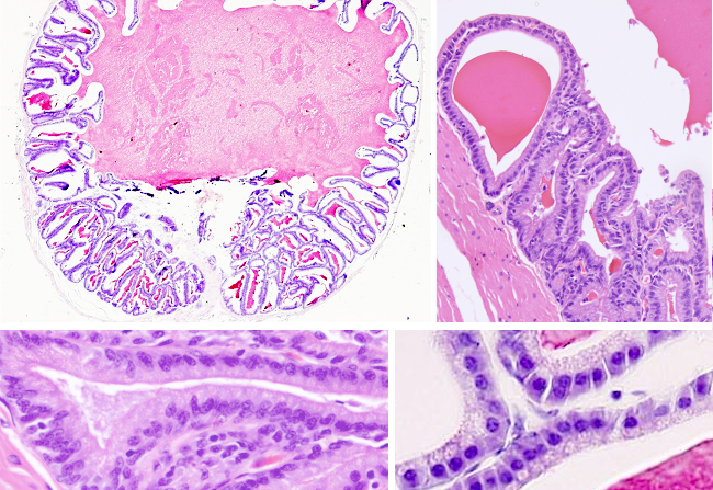
Animal organs. Reproductive.
SEMINAL VESICLE

Species: rat. (Rattus norvegicus; mammals).
Technique: paraffin embedding, section stained with haematoxylin and eosin.
Seminal vesicles are two tubular and irregular glands found above the prostate and behind the urinary bladder. Each seminal gland sends a short excretory duct that fuse with the ampule of the ductus deferens to form the ejaculatory duct. The walls of the seminal vesicle are made up of a characteristic highly folded mucosa. The folds are anastomosed and increase the secretory surface. There are primary folds that are in turn folded to form secondary folds, and sometimes tertiary folds can also be observed. The fusion of the epithelium of neighboring folds (anastomosis) produces irregular cavities connected between each other. In the image above, some cavities look isolated, but all the interior of the gland is actually connected.
The mucosa is made up of secretory epithelium and a non-well developed lamina propria of connective tissue. The organization of the epithelium is variable because pseudostratified and columnar cell arrangements can be found. Columnar epithelial cells show a basal elongated nucleus and many secretory vesicles in the apical cytoplasm. Lypofucsin granules have been observed. The secretory mechanism can be merocrine (exocytosis) or apocrine (the apical part of the cell is shed out). Secretory vesicles contain fructose for feeding sperm, together with other carbohydrates, amino acids, ascorbic acid and prostaglandins with unknown functions. It is interesting that flavins are also released because these molecules are used in forensics to detect the presence of semen since they emit a yellow color under the ultraviolet light, i.e., they are fluoresce molecules.
Below the mucosa, there is a well-developed smooth muscle layer with two sub-layers, one with obliquely oriented cells and another with longitudinally oriented cells. The muscle contraction happens during ejaculation, when seminal vesicles release the seminal fluid toward the ejaculatory ducts, which helps with the movement of the sperm along the urethra. Seminal vesicles were thought to be sperm storage compartments, but there is no sperm inside the vesicle cavities. They just release seminal fluid, which accounts for most of the sperm volume. Testosterone controls the production of seminal fluid.
During embryo development, seminal glands develop from the ductus deferens so that they inherit a similar tissue organization. However, seminal vesicles show a large cavity, a folded mucosa and a muscle layer (thinner than that of the ductus deferens). Aging reduces the seminal vesicle size and the mucosa becomes more flat.
