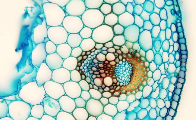
In this stem, the individual distribution of vascular bundles (Figure 1; they are separated from each other, and can be counted) is telling that it is primary growth. Vascular bundles are organized forming a circle (Figure 1), typical from dicots and gymnosperms. However, having tracheae in the xylem is a feature of dicot plants.

Epidermis is the protective tissue of this stem. It is an uniseriate layer of cellsshowing low cutinized cell walls. Parenchyma tissue is found underneath the epidermis. Parenchyma cells leave rather large intercellular spaces, particularly in the cortical region. Vascular bundles are organized as open collaterals and are protected by a bundle sheet of sclerenchyma with cell walls in redish because of lignin. Primary phloem is composed of sieve tubes with small companion cells. They are stained in blue because they have primary cell walls. The procambium is found between the primary phloem and the primary xylem, showing small cells stained in blue and more square in shape. In the primary xylem, the metaxylem consists of large trachea cells surrounded by sclerenchyma cells. Protoxylem is scarce and shows many parenchymatous cells.
Medullary region, or pith, has been reabsorbed and is empty in this stem, perhaps as an adaptation to wet environments.