

Cardiomyocytes, or cardiac muscle cells, form the cardiac muscle of the heart walls. They contract to pump the blood through the organism. Systole and diastole phases of the heart beating is the result of the contraction and relaxation of the cardiomyocytes, which means the contraction and relaxation of the heart ventricle walls.
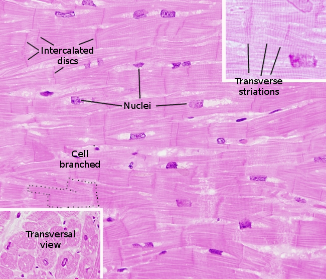
Cardiomyocytes are mono-nucleated cells, with the nucleus found in a central position of the cell (Figure 1). They are shorter (80 to 100 µm) and wider (about 15 µm) than skeletal muscle cells, and they are branched. Cardiomyocytes show striations with similar pattern to that of the skeletal muscle cells, with dark strips corresponding to the overlapping of the actin and myosin filaments, and clear strips that are only actin filaments. Myosin filaments are 1600 nm long and 10 nm thick, and are made up of about 400 myosin molecules. They form the A bands of the sarcomere. Myofibrils are no thicker than 1 µm.
In mammals, the plasma membrane of cardiomyocytes (or sarcolemma,) folds inward to form the t-tubules (transverse tubules). They are about 4 to 20 nm in diameter, larger than in the skeletal muscle cells, and are found at the level of the Z discs of the sarcomere. Another difference when compared with skeletal muscle cells is that, instead of triads, t-tubules form pairs with the endoplasmic reticulum. In addition, cardiomyocytes contain more mitochondria than the skeletal muscle cells.
Cardiomyocytes may increase and decrease their cellular thickness by hypertrophy and atrophy, respectively. This is caused by increasing or decreasing the number of cytoeskeletal myofibrils. However, cell proliferation is very rare.
Cardiomyocytes are anchored to one another by the so-called intercalated disks, which appear as dark strips in the plasma membrane of adjoining cells in heart histological samples. Intercalated disks are made up of cellular junctions, mainly desmosomes and adherent junctions (or fascia adherens), that keep a strong cohesion between cardiomyocytes. The cellular junctions of intercalated disks are the anchoring sites for the cardiomnyocyte cytoskeleton. Gap junctions are also observed between neighbor cardiomyocytes. They allow the electrical synchronization, i.e. contraction synchronization, of the heart muscle.
The rhythmic beating of the heart is modulated by the autonomous system, which adjust the frequency and intensity, but the rhythm is generated by special cardiomyocytes that work as pacemakers. That is why the heart muscle is regarded as involuntary muscle. The nerve fibers of the autonomous nervous system innervate these pacemaker cardiomyocytes to modify the rhythm. Not all cardiomyocytes are pacemakers, nor are they innervated by the autonomous system, because the gap junctions make possible the electrical coupling between all cardiomyocytes. The frequency of the heart beating is also regulated by some hormones.
Lardiomyocytes contain very low glycogen, so they cannot get much energy from glycolysis. The major part of the energy is provided by oxidative phosphorylation, with a high consumption of oxygen. Therefore, cellular damages are quickly produced when the oxygen supply is stopped.