

Animal tissues
Glandular epithelium
THYROID GLAND

Species: mouse (Mus musculus; mammal)
Technique: 8 μm paraffin section stained with PAS-haematoxylin.

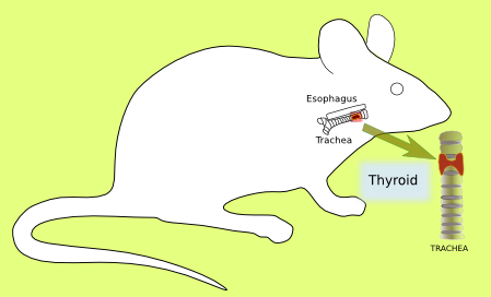
Thyroid, one of the largest endocrine glands of the body, is located at the front part of the neck, above the Adams apple, and shows a butterfly shape. Thyroid is divided in lobes, which are separated from each other by connective tissue. The blood vessels, into which hormones are released, run through this connective tissue. Thyroid lobes are composed of round follicles. Each follicle contains an acellular lumen surrounded by a simple columnar/cuboidal epithelium. The lumen is filled with colloid, a proteinaceous reservoir of thyroid hormone precursors, the most abundant one is thyroglobulin. The simple columnar/cuboidal epithelium is formed by follicular cells that synthesize and release two hormones: triiodothyronine (T3) and thyroxine (T4). Parafollicular cells are found among the follicles and release the calcitonin hormone.