Atlas of plant and animal histology

Español
Dark
Home / Animal tissues / Epithelium / Glandular / Intraepithelial cells
Site contents
The cell
Cell types
Animal tissues
Plant tissues
Animal organs
Plant organs
Histological techniques
Virtual microscopy

Animal tissue
Glandular epithelium


INTRAEPITHELIAL GLANDULAR CELLS

Pseudostratified epithelium
Intraepithelial gland cells
Cilium
Connective tissue
Labels
No labels
Training
Intraepithelial glandular cells
Organ: lung, respiratory tract, pseudostratified ciliated epithelium with intraepithelial glandular cells.
Species: pig (Sus scrofa domesticus; mammal)
Technique: 8 µm paraffin section stained with PAS-haematoxylin.

The image is from the respiratoty ducts.
Put the cursor over the mouse to see where the image comes from.

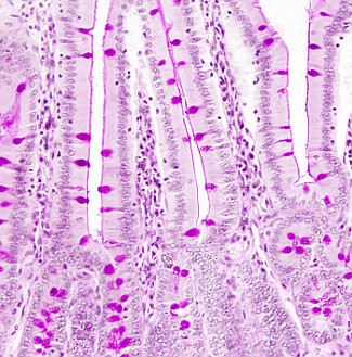
Globet cells (or caliciform cells) are intraepithelial glandular cells scattered in the covering epithelium of the intestine (small and large intestine), and larger respiratory ducts (trachea and bronchi). Globet cells resemble a calyx, with the apical part wider than the basal one. The nucleus is located in the basal part and the secretory vesicles are located toward the apical part (stained in redish in this image). The release of the secretory products is by merocrine secretion. After every secretion event, globet cells become thinner and begin to synthesize new molecules to be released in the next secretion event. The time between two releases is around 1 or 2 hours.

In some places, intraepithelial glandular cells form groups known as intraepithelial glands, which are completely included in the epithelium (see images below). Intraepithelial glands can be found in the nasal cavity, Eustachian ducts, urethra, eyelid and in the eye conjunctive.

Related pages
Small intestine
Large intestine

More images

Intraepithelial gland
Intraepithelial gland in the inner eyelid of a rat.
Glándula intraepitelial
Intraepithelial gland in the inner eyelid of a rat (magnification of the previous image).
Globet cells in the small intestine epithelium. These cells release mucus that covers the surface of the epithelium (note de red line lining the free epithelial surface).
Home / Animal tissues / Epithelium / Glandular / Intraepithelial cells