Animal tissues.
Epithelium.
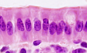
SIMPLE COLUMNAR

Species: human (Homo sapiens; mammal).
Technique: stainined with hematoxylin-eosin, 8 µm thick section, paraffin embedding.


The simple columnar epithelium is composed of cells that are taller than wider. They show a rounded nucleus located in the basal part of the cell (see also figure 1). The apical surface of the cell frequently shows cilia that, thanks to their movement, can transport substances over the surface of the epithelium. Ciliated simple columnar epithelium can be found in the uterine tube. Microvilli may also be present in the apical surface of simple columnar epithelium, as in the intestine and gall bladder epithelia. Microvilli increase the absorptive surface of the epithelium. Epithelial cells are anchored to the basal lamina by hemidesmosomes. There are tight junctions and adherent junctions close to the apical surface that seal the intercellular space, preventing substances to cross the epithelium through the intercellular space. The lateral wall usually show many desmosomes that keep the structural integrity of the epithelial layer. The latero-basal membranes are highly folded and form interdigitations with membranes of neighbor cells.

Más imágenes