Animal tissues.
Covering epithelium.
TRANSITIONAL EPITHELIUM

Species: mouse (Mus musculus; mammal).
Technique: haematoxylin-eosin, 8 µm thick section, paraffin embedding.
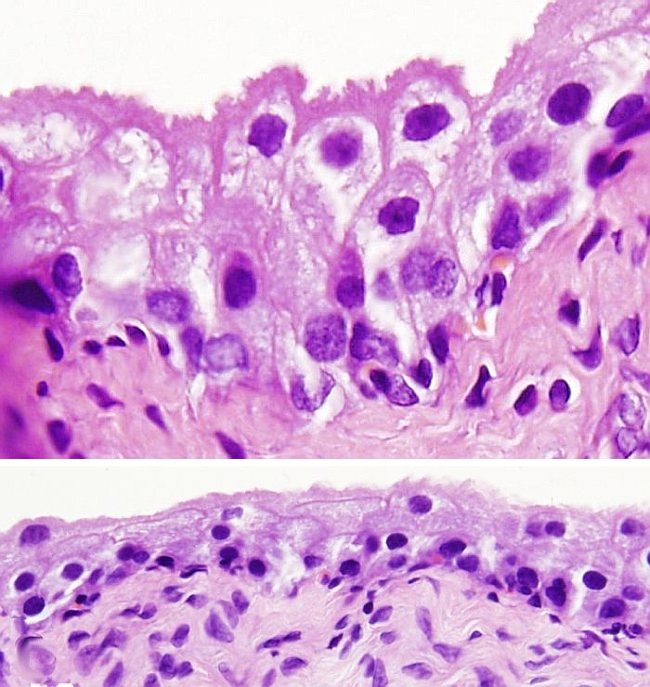
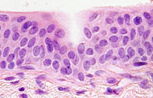
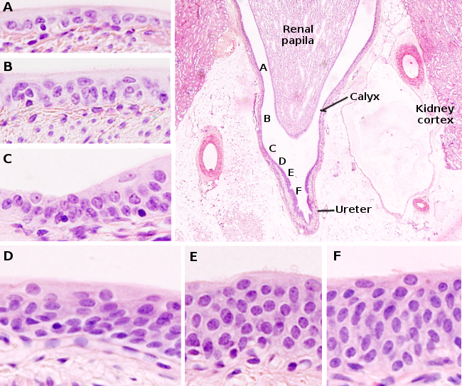
It was thought that transitional epithelium was an intermediate type of epithelium, a “transition”, between the stratified squamous epithelium and the stratified columnar epithelium. That is why the name transitional. But it is not. The transitional epithelium is a stratified epithelium with a variable number of cell layers (2 to 6), which are irregular in thickness and in the position of the nuclei (see also figure 1). The transitional epithelium is also called urothelium because it lines urinary ducts, such as renal calyxes (2 cell layers), urethers (3 to 5 cell layers), urethra (4 to 5 cell layers) and urinary bladder (up to 6 cell layers). It is not very permeable to salts and water, and it acts as an osmotic barrier between the urine and tissues. Actually, the transitional epithelium is a better barrier than the epidermis.

Three types of cells are distinguishible in the transitional epithelium.
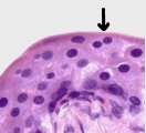
The superficial cells are polyhedral cells that change their morphology according to the amount of urine in the urinary compartment: they are flat (stretched) when the duct or urinary bladder are empty, and rounded (relaxed) when they are empty. These cells form the impermeable barrier to diffusion between urine and blood. The superficial cells have plate-like structures, separate by membrane regions known as hinges. The plates contain a protein called uroplaquin. The tight junctions (adhesion complexes) between superficial cells also contribute to the impermeability, as well as a glycan layer in the plasma membrane and a plasma membrane with a particular composition. The high-stretching capacity of the transitional epithelium, after filling the urinary compartments, is because these superficial cells can be flattened. In a relaxing state, the apical surface of the epithelium looks wrinkled because the plates and hinges get folded. Depending on the species, some cells of the superficial layer may be binucleated (Figure 2) and the nuclei may be polyploids.

Under the superficial layer, there are one or two layers of intermediate cells ( 5 or 6 layers in humans). These cells are from fusiform to columnar, with a nucleus, and connected between one another through desmosomes.
The basal cells are arranged in a one thick-cell layer, in contact with the basal lamina. They are tightly packaged and smaller than those of the upper layers. The adult stem cells that proliferate and differentiate to replace cells of the upper layers are found in this layer. As they detach from the basal layer and migrate toward the superficial layer, they increase in size and differentiated. The proliferative activity of the transitional epithelium is low under normal conditions, but it may be increased after injuries, when it shows a strong regenerative capability. For example, when superficial cells are damaged, intermediate cells quickly differentiate to replace the superficial lost cells. It has been proposed that all cells of the transitional epithelium send processes that make physical contact with the basal lamina, at least in some species.
Bibliography
Arrighi S. 2015. The urothelium: Anatomy, review of the literature, perspectives for veterinary medicine. Annals of anatomy. 198: 73-82.
Más imágenes


