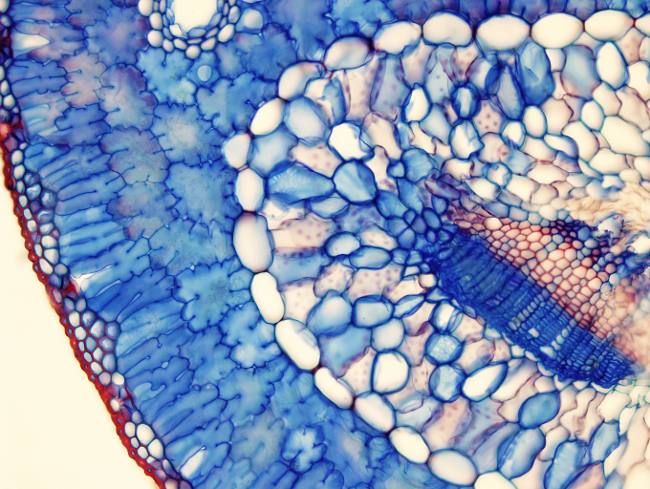
Pine leaf or acicular show an epidermis with highly lignified cells. Under the epidermis, there are 2 or 3 layers of cells called hipodermis, which also have lignified secondary walls and look like macrosclereids. Both adaxial and abaxial layers have stomata. They are sunken stomata because their occlusive cells are below the level of the epidermis. The Mesophyll is homogeneous in this leaf and all phosynthetic parenchyma cells look like palisade parenchyma having little intercellular spaces. A layer that looks like an endodermis wraps the transfusion tissue and the vascular bundle. The homogeneous appearance of the xylem is because it is made up of tracheids, but there are no tracheae, and this is a feature of gymnosperm xylem. Phloem is composed of sieve cells tightly piled and are difficult to discern from the companion parenchyma cells. The transfusion tissue contains cells with easy-visible pores, which are structures for intercellular communication. In this image, they can be observed as faint brown discs. Cells having the pores may be tracheids intermingled with storing parenchyma cells, that have a more intense stained blue cell wall.
