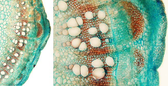
The picture above is from a secondary stem since there is no vascular bundles, that is, vascular bundles with interfascicular parenchyma have disappeared. It is a dicot plant because it contains tracheae in the xylem. Although secondary xylem and secondary phloem are clearly visible, this stem is starting the secondary growth because there is no periderm yet (see Figure 1).

The epidermis of the stem of the large image is an uniseriate layer containing non-cutinized cells. Under the epidermis, there is a cortical parenchyma with tightly packaged cells and some scattered sclerenchyma fibers, which were formed during the primary growth of the stem as part of the bundle sheet surrounding the primary vascular bundles. Cells of the primary phloem (metaphloem) are compressed below the sclerenchyma cells. In the secondary phloem, two systems can be observed: the vertical or axial system (parallel to mayor axis of the stem), found under the primary phloem, and the horizontal system formed by rays of parenchyma (arranged perpendicularly to the major axis of the stem). The rays of parenchyma of the secondary phloem are continuous with the rays of parenchyma of the secondary xylem (xylem rays). In the secondary xylem, large tracheae can be observed intersperse with sclerenchyma fibers. The primary xylem, protruding in the medullary parenchyma, is made up of smaller tracheae and parenchyma cells. The vascular cambium is found between the secondary phloem and the secondary xylem as a continuous line, that in tridimensional view is actually a cylinder.
