

Animal tissues.
Glandular epithelium.
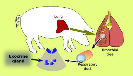
EXOCRINE GLAND

Organ: lung, respiratory ducts, exocrine gland
Species: pig (Sus scrofa domesticus; mammal)
Technique: PAS-haematoxylin, 8 µm thick section, paraffin embedding.
Species: pig (Sus scrofa domesticus; mammal)
Technique: PAS-haematoxylin, 8 µm thick section, paraffin embedding.

 This image is from respiratory ducts.
Put the cursor over the mouse to see where the image comes from.
Exocrine glands show an excretory duct, which connects the secretory units (structures in red in the image above) with the free surface of the epithelium. The morphology and arrangement of both secretory and excretory parts result in a wide variety of exocrine glands. Compound exocrine glands show branched excretory ducts and simple exocrine glands are not branched and have one secretory unit. Regarding the secretory units, exocrine glands are known as tubular (tubuloacinar), acinar, or alveolar, depending on the organization and morphology of the secretory cells (see table).
More images


