1. Morphology
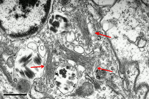
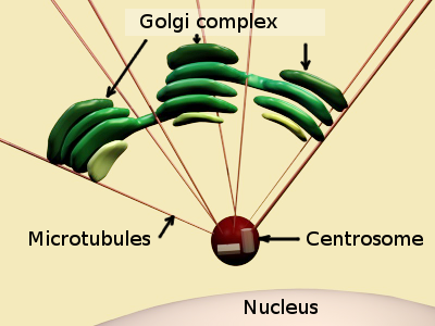
The Golgi apparatus was discovered by Camillo Golgi in 1889, and studied at electron microscopy by Dalto and Felix (1954). In animal cells, the Golgi apparatus is commonly found near the centrosome, which in turn it is commonly found close to the nucleus. Therefore, the Golgi apparatus location depends on microtubules. Th Golgi apparatus is made up of flat cisterns piled in several stacks. Each stack is referred to as dictyosome (Figures 1). Several stacks are present in one cell and some cisterns connect neighboring stacks (Figure 2). The Golgi apparatus includes all the stacks and their lateral connections.


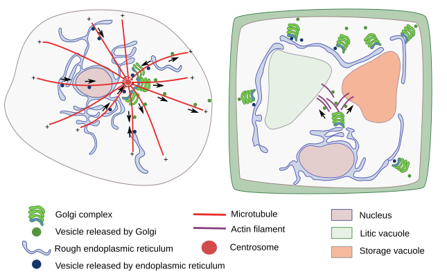
In animal cells, there is a fibrous protein matrix among the Golgi apparatus cisterns that may help to maintain the organelle structure. However, it has been shown that the structural integrity of the Golgi apparatus strongly relies on microtubule organization (Figure 3).

In plant cells, where no centrosome is present, there are small stacks of cisterns, or even individual cisterns, dispersed throughout the cytoplasm (Figure 3). The Golgi apparatus looks like divided in many units distributed through the cytoplasm. Individual cisterns or stacks are moved thanks to actin filaments. In some eukaryote cells, such as yeast, there are not even cisterns similar to those found in plant and animal cells.
2. Organization
The Golgi apparatus is a polarized organelle. The cistern stacks are organized in three domains: cis, intermediate and trans domain (Figure 4). Intermediate cisterns are found between cis and trans domains. In the cis domain, there is an ongoing addition of new material coming from the endoplasmic reticulum via the ERGIC (endoplasmic reticulum Golgi intermediate compartment) compartment. In the trans domain, there is a tubulo-vesicular arrangement of the membranes, referred to as TGN (trans Golgi network), where molecules are sorted into vesicles and tubules in their way toward other cell compartments. Thus, the molecule journey through the Golgi apparatus starts in the cis cisterns, goes through intermediate cisterns, and ends up in the trans domain. The Golgi apparatus is particularly well-developed in those cells showing an intense secretory activity.

3. Golgi apparatus models
a) Cisternal maturation model (Figure 5). It proposes that ERGIC bodies loaded with endoplasmic reticulum material get fused between each other to form the first cistern at the Golgi cis domain. Then, this cistern moves toward the trans domain, where it is eventually split in vesicles. The internal material of the cistern is progressively processed during this journey, i.e., there is a maturation of the cistern as it moves through the Golgi apparatus. Currently, this is the most accepted model because it can explain some observations that do not fit in other models.

b) Connection by tubules. Tubular connections between adjoining cisterns have been observed at electron microscopy. These connections are like bridges that appear to be transient and depending on the material to be processed. This proposal is not conflicting with the other model, and both processes may work concurrently.
4. Functions
a) The Golgi apparatus is a major glycosylation center of the cell. Many carbohydrates that are part of glycoproteins, proteoglycans, glycolipids and other polysaccharides, such as hemicellulose in plants, are assembled and modified in the Golgi apparatus. Sialic acid is a carbohydrate specifically added in this organelle. Carbohydrates are linked through a particular type of binding known as O-type glycosylation. Sulfation of proteoglycans also happens in the Golgi apparatus, as well as phosphorylation, palmitoylation, methylation, and other chemical modifications. In plants, the glycosylation function of the Golgi apparatus is essential because many glycoconjugates are part of the cell wall. However, cellulose is synthesized at the cell membrane. All chemical reactions involving carbohydrates are carried out by glycosyltransferases and glycosidases. These enzymes are specifically distributed along the cis-trans axis of the Golgi apparatus.
b) The synthesis of sphingomyelins and glycosphingolipids is finished in the Golgi apparatus.
c) The Golgi apparatus is a center for sorting and shipping molecules coming from the endoplasmic reticulum or synthesized in the Golgi apparatus itself. Once processed, molecules are sorted into different types of vesicles and targeted to other cell compartments. In the trans domain, the TGN is the structure where this process takes place. Some vesicles are moved to and fused with the plasma membrane, a process known as exocytosis. There are two types of exocytosis: constitutive and regulated (see next page). Furthermore, there are vesicles leaving the trans domain targeted to late endosomes/multivesicular bodies, or to vacuoles in plants. The trans domain is also a target for other vesicles coming from the endosomes, for recycling. There is also a bidirectional communication between the Golgi apparatus and the endoplasmic reticulum.
-
Bibliography ↷
-
Bibliography
Farquhar, MG, Palade GE. 1981. The Golgi apparatus (Complex) - (1954-1981) - from artifact to center stage. The journal of cell biology. 91: 77s-173s.
Glick, BS, Luni A. 2011. Models for Golgi traffick: a critical assessment. Cold Spring Harbor perspectives in biology. 3: a005215.
Hellicar J, Stevenson NL, Stephens DJ, Lowe M. 2022, Supply chain logistics – the role of the Golgi complex in extracellular matrix production and maintenance. Journal of cell sciences. 135: jcs258879. doi:10.1242/jcs.258879.
Vildanova MS, Wang W, Smirnova EA. 2015. Specific organization of Golgi apparatus in plant cells. Biochemistry (Moscow). 79: 894-906.
Witkos TM, Lowe M. 2017. Recognition and tethering of transport vesicles at the Golgi apparatus. Current opinion in cell biology. 47:16–23.
-
 Fron reticulum to Golgi