Microtubules are a component of the cytoskeleton performing many functions. They contribute to the spatial organization of organelles, work as tracks for vesicular trafficking, are needed during cell division by forming the mitotic spindle, participate in cell movement, contribute to cell polarization in some cell types, and are an essential component of cilia and flagella.
1. Structure
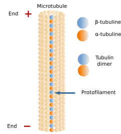
Microtubules are long and relatively stiff tubules (Figures 1). Their walls are made up of many dimers of globular proteins: α- and β-tubulin (Figure 2), which are lined up in long rows known as protofilaments. A microtubule is usually composed of 13 protofilaments. Microtubules are polarized structures, with a minus and a plus end, and are dynamic by polymerization and depolymerization. New tubulin dimers are mainly added to the plus end, where the growth of the microtubule usually happenss. In the minus end, depolymerization prevails over polymerization. Tubulin dimers alternate between the ends of the microtubule and the cytosol, which is a dynamic behavior, allowing the reorganization of the microtubule cell net as needed.


2. Dynamic instability
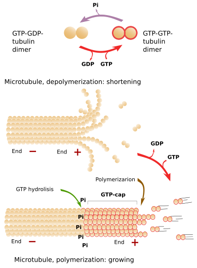
The addition of new tubulin dimers to the plus end makes the microtubule grow in length. The growth is stopped from time to time, and growth periods alternate with shrinkage periods. Depolymerization is sometimes so strong that the complete microtubule may disappear. However, it is more frequent a new polymerization (growing) period. The alternation between polymerization and depolymerization of microtubules is known as dynamic instability.
Free tubulin dimers are linked to two GTP molecules. Some time later, after the joining of a tubulin dimer to the plus end of a microtubule, one GTP is hydrolyzed to ADP (Figure 3). If the rate of adding GTP-GTP-tubulin dimers to the plus end is faster than the hydrolysis rate, there will always be a group of tubulin dimers with GTP-GTP in the plus end, which is known as GTP-cap. GTP-cap stabilizes the plus end of the microtubule and boosts polymerization. If the addition rate of new GTP-GTP-tubulin dimers is low, the hydrolysis rate may overcome the polymerization speed. This means that there are GTP-GDP-tubulin dimers at the plus end, which makes protofilaments weakly adhere to one another. In this situation, massive depolymerization starts. If the plus end is stabilized by external elements, the microtubule grows again (Figure 3).

3. MAPs
There are microtubules associated proteins (MAPs) controlling the organization, stability, growth, and other aspects of microtubule behavior. MAPs may interact with the microtubule plus end affecting the dynamic instability, either by boosting grow or depolymerization. MAPs also allow microtubules to interact with other cellular elements, such as organelles or cytosolic molecules, such as other cytoskeleton components. Some substances that affect polymerization or depolymerization of microtubules. For example, colchicine inhibits microtubule growth, whereas taxol strongly attachs to microtubules, preventing depolymerization.
4. MTOCs
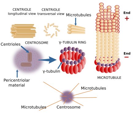
New microtubules are formed from scratch by nucleation. In the cell, there are molecular structures known as microtubule organizing centers (MTOCs), where microtubules are nucleated. They contain γ-tubulin rings that work as templates for new microtubules.
The centrosome is the major MTOC in animal cells (Figure 4). It is the main responsible for the number, localization and spatial organization of microtubules. In most animal cells, during the G1 and G0 phases of the cell cycle, there is one centrosome per cell found near the nucleus. However, megakaryocytes contains multiple centrosomes, and muscle fibers lack centrosomes. The centrosome consists of two components: a couple of orthogonally oriented centrioles and a surrounding pericentriolar material.

There are many γ-tubulin molecules in the pericentriolar material arranged in rings, known as γ-tubulin rings, that nucleate microtubules. Plant cells lack centrioles and do not form typical centrosomes, but they have γ-tubulin rings associated with the nuclear envelope, blepharoplasts, and are scattered through the cytoplasm. The main MTOC in yeasts is the polar body, which is inserted into the nuclear envelope. There are other microtubule nucleators such as chromosomes, which can form a mitotic spindle without centrosomes. The cisterns of the Golgi apparatus nucleate microtubules that help to maintain the general organization of the organelle.
5. Function
Microtubules are classified into two types: stable microtubules, found in cilia and flagella, and dynamic microtubules, found in the cytosol. Cytosolic microtubules, besides their role in the mitotic spindle formation and chromosome segregation, are also involved in the internal movement of organelles such as mitochondria, lysosomes, pigment inclusions, lipid drops, etcetera. This transport is carried out by motor proteins that can move along the microtubules, pulling cargoes. Kinesins and dyneins are the two families of motor proteins associated with microtubules. Despite transporting, motor proteins help to maintain the location and morphology of cell organelles, such as the Golgi apparatus and endoplasmic reticulum.
Cilia and flagella are filiform cellular structures protruding from the cell surface and contain microtubules. Cilia are shorter than flagella and more numerous, and their movements propel the liquid parallel to the cell surface. Flagella move the surrounding liquid perpendicularly to the cell surface. Both have a central structure of microtubules known as axoneme. The axoneme is composed of 9 outer pairs of microtubules, plus a central pair of microtubules. The axonem is polymerized from a basal body, which is similar to a centriole (9x3+0). The outer pairs of microtubules of the axoneme are connected to each other by nexin proteins, whereas protein spokes connect the outer pairs with the central pair. The motor protein dynein is located between the outer pairs. Dynein is involved in the movement of cilia and flagella. Some types of cilia are known as primary cilia. They lack the central pair of microtubules and are sensory structures.
 Actin filaments