
Glossary: M
A B C D E F G H I J K L M N O P Q R S T U V W X Y Z
Mast cell: see mastocyte.
Macrophage: it is a type of cell derived from monocytes and found in the connective tissue. The main function of macrophages is to phagocyte particles: those recognized as foreign particles by the immune system, such as virus, bacteria and other pathogens, as well as particles resulting from death cells. Macrophages contain a well-developed lysosomal system for molecular hydrolysis.
Main olfactory epithelium: it is a pseudostratified epithelium found in the upper part of the nasal cavity. In humans, it is about 1 cm2. The main cell type of the main olfactory epithelium is the olfactory receptor, a specialized neuron type. Olfactory receptors are able to recognize odoriferous molecules and send the information to the encephalon for sensory processing and integration.
Malleus: (or hammer) it is a small bone, that together with the incus and stapes, form the middle ear ossicles. The malleus in found in the tympanic cavity, in contact with the tympanic membrane and with the incus. It transmits the tympanic membrane vibrations to the incus.
 .
.Margin: (in plant leaves) it is the edge of the leaf. The leaf margin may show a wide variety of morphologies: lobated, serrated, dentate, entire, etcetera.
 .
.Mastocyte: it is a type of cell found in the connective tissue, mainly in the skin, digestive tract and respiratory tract. Mastocytes are very large cells involved in the allergic and anti-inflammatory responses. They release histamine and heparin. In the mastocyte cytoplasm there are many granules that store these and other substances.


 .
. ,
,Mechanoreceptor: it is a sensory receptor found in animals that detects physical changes like pressure, movement of the body or movement of an internal liquid. Mechanoreceptors are the Merkel discs, some free nerve terminals and the encapsulated receptors. In the middle ear, the Corti organ is a mechanoreceptor.
Megakayriocyte: it is a type of cell found in the bone marrow. Megakaryocytes are part of the hematopoietic system. They are large polynucleated cells that produce the blood platelets (or thromobocytes).
Megaphyll: (in plants) it is a type of leaf showing a complex pattern of branched veins. Megaphyll leaves are typical of flowering plants. Microphyll are leaves with simple vein organization.
Megaspore: (in plants; also called macrospore) it is the cell that results after meiosis in the ovule of flowers. Female gametophyte is formed from the megaspore by a process known as megagametogenesis.
 .
.Megasporogenesis: (in plants) it is the process of formation of the megaspore, or egg, in the ovule of flowers.
Meissner corpuscle: it is a type of encapsulated receptor found in the dermis, usually in the dermal papillae of regions lacking hair follicles. The capsule is made up of layers of connective tissue cells, whith nerve terminals among them. A mechanical stimulus leads to a separation of the layers from one another, resulting in a modification of the nerve membrane. Meissner corpuscles show fast adaptation to stimuli, so that even if the stimulus is sustained the nerve terminal stop sending information. However, they are very useful to detect the movement of objects over the skin. That is, they are able to inform of the spatial movement component of the stimuli, as well as texture. Meissner corpuscle are abundant in the regions requiring a high discrimination of mechanical stimuli like fingertips and lips. The nerve terminal is organized as a spiral. Those Meissner corpuscles found in the genitals and nipples are known as genital corpuscles.
Melanin: it is a protein synthesized and released by melanocytes, cells found in the stratum basale of the epidermis. Melanin provides the dark color to the skin and protects against the ultraviolet radiation.
Melanocyte: it is a type of cell found in the stratum basale of the epidermis, among the keratinocytes. Melanocytes synthesize and release melanin, protein that provides the dark color to the skin and protects against the ultraviolet radiation.
Membranous labyrinth: it is the set of membranes found inside the bone labyrinth. Membranous labyrinth lines the sacule, utricle, semicircular canals and cochlea. There are sensory cells in the membranous labyrinth that respond to head movement, such as acceleration, position, as well as sensory cells of the Corti's organ for sensing sound.
 .
.Meristem: (in plants) it is a group of undifferentiated cells found in different parts of the body plant. Meristems cells can proliferate and differentiate into all the cell types of the plant. There are different types of meristems. Primary meristems allow the growth in length of plants and are usually found at the tips of stems and roots, as well as in the internode regions. Secondary meristems are in charge of the growth in thickness in those plants and organs with secondary grow, and are found in the stems and roots.
 ,
Root primary meristem
,
Root primary meristem


 ,
Shoot primary meristem
,
Shoot primary meristem

 ,
Vascular cambium
,
Vascular cambium


 ,
Cork cambium
,
Cork cambium
 .
.
Meristematic cell: (in plants) it is a type of undifferentiated cell found in plant meristems. Meristematic cells differentiate into functional cells to form new tissues that leads to organs and plant growing. They can also give rise to new meristematic cells by mitosis, so that there is always a population of undifferentiated cells in plant meristems.
 ,
,Merkel cell: it is a type of cell found in the epidermis of animals. Merkel cells perform a sensory role as mechanoreceptor.
Merkel's disk: it is a type of mechanosensory receptor. Merkel's disks are made up of free nerve terminal organized as a cup that encloses a Merkel cell of the epidermis. Merkel's disks are very sensitive and show slow adaptation, so they inform about long-lasting stimuli. They are abundant in the fingertips and lips, although they are found in the skin of the rest of the body.
Merocrine secretion: it is a type of secretion where the secretory cells release substances by exocytosis.
 .
.Mesenchymal cell: (in animals) it is a type of cell abundant in embryo tissues, but it is also present in adults. Mesenchymal cells are undifferentiated cells that give rise to many cell types. For instance, most connective tissue cells. They show star-like or fusiform shapes and may develop a high proliferative rate. The majority of mesenchymal cells derive from mesoderm and, in some parts of the body, from neural crests (endoderm).
 .
.Mesenchymal tissue: it is a type of connective tissue consisting of undifferentiated cells and an amorphous extracellular matrix lacking collagen fibers and elastic fibers. In the embryo, the mesenchymal tissue differentiates into other tissues, such as other connective proper tissues, cartilage, and bone.
Mesentery: it is a fold of the peritoneum that connects parts of the small intestine and stomach with the abdominal wall.
Mesoderm: it is one of the three germ layers found in the embryo at the end of gastrulation. Mesoderm cells differentiate in many adult structures in animals, such as most muscles, bone skeleton, urogenital system, blood, and many others.
Mesophyll: (in plants) it is a type of parenchyma found in leaves. Mesophyll locate between the upper and bottom epidermal layers of leaves. The majority of mesophyll is photosynthetic parenchyma.

 .
. .
.Mesothelium: it is a simple squamous epithelium the delimits the body cavities. The thorax cavity is limited by pleura (mesthelium), the abdominal cavity by peritoneum (mesothelium), the heart is in the cardiac cavity delimited by mesothelium too. Mesothelium also delimits the synovial cavity at joints and can be found in the inner reproductive organs.
Metaphloem: (in plants) it is the primary phloem that appears after protophloem during growing of the plant organs. Metaphloem is the functional phloem in monocots and dicots during primary growth. It is substituted by secondary phloem in those organs with secondary growth. Metaphloem is composed of sieve elements, companion and parenchyma cells.
 ,
, .
.Metaxylem: (in plants) it is the primary xylem that develops after the protoxylem, during the primary grow of plant organs. Metaxylem is substituted by secondary xylem in organs with secondary growth. In organs without secondary grow, the metaxylem is the functional xylem. Metaxylem is composed of vessels elements, parenchyma cells, sclerenchyma cells and sclereids.
 .
. ,
Dicot primary stem
,
Dicot primary stem
 ,
Monocot primary stem
,
Monocot primary stem
 ,
Gymnosperm secondary stem.
,
Gymnosperm secondary stem.
Microphyll: (in plants) it is a type of leaf with a simple vein system organization. Micropyll leaves are present in the ferns. Those leaves with complex vascularization are called magaphyll, for instance in angiosperms.
Micropyle: (in plants) small passage in the female gametophyte that allows the nuclei of the pollinic tube to get in contact with female nuclei. Thus, fertilization takes places. Later in the seed, the muicropyle is a small pore that allows the water to enter the seed and starts germination.
 .
.Microspore: (in plants) it is a type of cell that is the result of meiosis of the anther cells of stamens. Microspores divide and differentiate into the male gametophyte by a process known as microgametogenesis.
 .
.Microsporogenesis: (in plants) it is the process of microspore formation in the interior of the anther of the stamens.
Microvilli: they are densely packaged filiform expansions of the apical surface of some epithelial cells, like the intestine epithelium. An epithelial cell may contain hundreds them. Each microvillus have the same length (about 7 µm), resulting in a brush-like structure in the apical domain of the epithelial cell. They contain an actin scaffold. Microvilli increase the surface of the cell, which results in a higher absorption capacity by the eptihelium.


 ,
, .
. .
.Middle ear: it is a part of the ear, just next to the external auditory canal. The middle ear is a cavity, known as tympanic cavity, situated in the temporal bone of the skull. It is separated from the external auditory canal by the tympanic membrane. In the tympanic cavity, there are the three ossicles: the malleus, incus and stapes, and the muscles responsible for their movement. The Eustachian canal also belong to the middle ear, which connects the tympanic cavity with the pharynx.

Middle lamella: (in plants) it is the outermost layer of the cell wall, shared by adjoining cells. Middle lamella is the first layer of the cell wall to be synthesized during the cellular division. The aspect of the middle lamella is amorphous and contains many pectic substances.
 ,
,
 .
.Mitosis: it is the phase of the cell cycle where chromatin condenses into chromosomes and a proportional segregation of chromosomes between the two daughter cells. Mitosis includes prophase, metaphase, anaphase and telophase.
 ,
, .
.Mitral cell: it is a type of cell found in the olfactory bulb of animals. Mitral cells extend their dendrites into the olfactory glomeruli. They receive direct information from the olfactory receptors, and send the processed information to deep regions of the encephalon.
Mixed nerve: it is a nerve made up of both, axons that carry information from the central nervous system to the rest of the body and axons that carry information from the body to the central nervous system. That is, mixed nerves contain afferent and efferent axons.
Mixed secretion: it is a type of secretion where different secretory cells synthesize different substances that are released together. For instance, some salivary glands contain serous and a mucous secretory components.
Monocot: (in plants) it is a group of plants with seeds having only one cotyledon. Cotylendons are embryionary leaves that develop during germination. Monocot flowers usually show three petals or multiple of 3, and leaves show parallel oriented veins. The vascular bundles are scattered in the stem and the root do not branch from a main root, but all grow from the stem, that is, they show a fasciculate root system.
Monocyte: it is a type of non-granular leukocyte. Monocytes are the largest cells of the blood. They show an indented nucleus and abundant cytoplasm. Its cytoplasm and nucleus are stained lighter than those of other leukocytes when common staining methods are used. Monocytes are the direct precursors of macrophages.

Monoecious: (in plants; or monoicous) it is a type of plant having both male and female gametophytes.
Motor protein: it is a type of protein that transport cargoes along microtubules and actin filaments, by consuming ATP. Motor proteins are involved in mitosis, cytokinesis, ciliary movement, muscle contraction, intracellular transport, location and organization of organelles, and in many other functions. There are several families of motor proteins: dyneins and kinesins are associated with microtubules, and myosins are associated with actin filaments.
Mucilage: (in plants) it is a glucidic substance found in the aquiferous parenchyma that is able to fetch and store water.
 .
.Mucous accinus: it is a type of secretory unit found in some exocrine glands. Acini mucous cells show clear cytoplasm when stained with common techniques, like haematoxylin and eosin. The nuclei of these cells are flattened and found near the basal membrane. Acini mucous cells release water and mucopolysaccharides.
 ,
, .
. .
.Mucous gland: it is a type of gland characterized by releasing mucus, a substance composed of water, glycosaminoglycans (also known as mucopolysaccharides), salts and some proteins. The main function of mucous glands is lubricate the body surfaces. Calyciform cells of the respiratory tract and sublingual salivary glands are examples of mucous glands.
 ,
, ,
,Mucous secretion: it is a type of secretion largely consisting in glycosaminoglycans, some proteins, salts and water.
Mucous connective tissue: see gelatinous tissue.
Multilocular adipocyte: it is a type of adipocyte that forms the brown fat. The lipid droplets of these adipocytes are small and scattered in the cytoplasm. That is why the name multilocular.
 .
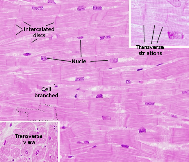
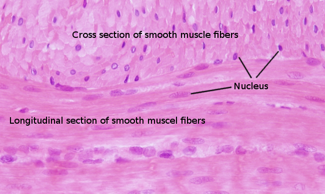
.Muscle cells: it is a type of cell that forms the animal muscles. The main feature of muscle cells is the ability to shorten the cell length, i.e., contraction. There are three types of muscle cells. Skeletal muscle cells, cardiac muscle cells and smooth muscle cells. Skeletal muscle cells form the bone-attached muscles, tongue and upper esophagus muscles. They are very long and multinucleated cells showing many striations. Skeletal muscle cells are under voluntary control. Cardiac muscle cells form the heart walls. They are branched, mononucleated and striated cells under involuntary control. Smooth muscle cells are mononucleated, do not show striations and are shorter than skeletal muscle cells. These cells form the walls of the digestive ducts and stomach, walls of blood vessels, and other organs that are under involuntary control.

 ,
, ,
, ,
,Muscle fascicle: it is a bundle of muscle cells within a muscle wrapped by a layer of connective tissue called perimisium. A muscle is made up of several muscle fascicles, which are better distinguished in skeletal muscles.
 ,
,Muscle tissue: it is one of the four fundamental tissues of animals. The main function of the muscle tissue is to produce movement by contracting the muscle cells. There are four types of muscle tissue: cardiac (heart walls), skeletal (voluntary body muscles) and smooth (involuntary visceral muscles).
Muscular artery: it is middle size artery with histological organization between elastic arteries and arterioles. The diameter ranges from 0.1 to 10 mm. Thus, larger muscular arteries are similar to elastic arteries, and the smaller ones are similar to arterioles. They do no show a particular feature compared to elastic arteries and arterioles. They contain less elastic fibers and more density of smooth muscle cells than elastic arteries.
Muscular vein: it is a type of vein found after the postcapillary veins. Muscular veins are about 1 mm in diameter and show well-developed walls with a visible tunica media showing one or two layers of smooth muscle cells. They also show a thin tunica adventitia.
Myelin: it is a sheath that wraps axons. Myelin is made up of many layers of glia cell plasma membrane. These glial cells are oligodendrocytes in the central nervous system and Schwann's cell in the peripheral nervous system. Myelin is na insulating layer that makes possible a very fast conduction of action potential along axons. Axons with myelin are referred as myelinic fibers or myelinic axons.
 .
. .
. ,
,Myelinic nerve: it is a nerve with the axons wrapped by myelin sheaths.
Myocardium: it is one of the three layers that form the ventricle and auricle walls of the heart. Myocardium is made up of cardiac muscle cells (cardiomyocytes) and some connective tissue. Ventricle wall is thicker than auricle wall, and consists of two layers: inner and outer. Cardiomyocytes are oriented in spiral in the outer layer, whereas in the inner one they are oriented circularly around the ventricle.
 .
.Myocyte: it is how mature muscle cells are also named. The heart mature muscle cells are called cardiomyocyte, and the skeletal muscle cells are also known as muscle fibers.

 ,
,Myoepithelial celll: it is a type of cell found around the secretory units of some exocrine glands. Myoepithelial cells may reduce their length by contraction, which drives the secretory products from gland secretory units to excretory ducts.